

Colposcopy and Treatment of Cervical Intraepithelial Neoplasia - RHO
Colposcopy and Treatment of Cervical Intraepithelial Neoplasia - RHO
Colposcopy and Treatment of Cervical Intraepithelial Neoplasia - RHO

Create successful ePaper yourself
Turn your PDF publications into a flip-book with our unique Google optimized e-Paper software.
Chapter 13<br />
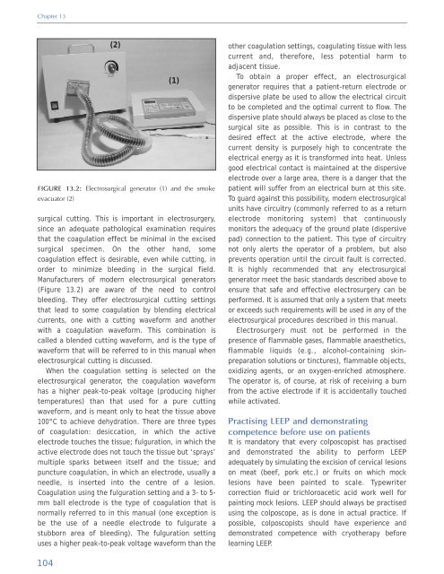
(2)<br />
(1)<br />
FIGURE 13.2: Electrosurgical generator (1) <strong>and</strong> the smoke<br />
evacuator (2)<br />
surgical cutting. This is important in electrosurgery,<br />
since an adequate pathological examination requires<br />
that the coagulation effect be minimal in the excised<br />
surgical specimen. On the other h<strong>and</strong>, some<br />
coagulation effect is desirable, even while cutting, in<br />
order to minimize bleeding in the surgical field.<br />
Manufacturers <strong>of</strong> modern electrosurgical generators<br />
(Figure 13.2) are aware <strong>of</strong> the need to control<br />
bleeding. They <strong>of</strong>fer electrosurgical cutting settings<br />
that lead to some coagulation by blending electrical<br />
currents, one with a cutting waveform <strong>and</strong> another<br />
with a coagulation waveform. This combination is<br />
called a blended cutting waveform, <strong>and</strong> is the type <strong>of</strong><br />
waveform that will be referred to in this manual when<br />
electrosurgical cutting is discussed.<br />
When the coagulation setting is selected on the<br />
electrosurgical generator, the coagulation waveform<br />
has a higher peak-to-peak voltage (producing higher<br />
temperatures) than that used for a pure cutting<br />
waveform, <strong>and</strong> is meant only to heat the tissue above<br />
100°C to achieve dehydration. There are three types<br />
<strong>of</strong> coagulation: desiccation, in which the active<br />
electrode touches the tissue; fulguration, in which the<br />
active electrode does not touch the tissue but ‘sprays’<br />
multiple sparks between itself <strong>and</strong> the tissue; <strong>and</strong><br />
puncture coagulation, in which an electrode, usually a<br />
needle, is inserted into the centre <strong>of</strong> a lesion.<br />
Coagulation using the fulguration setting <strong>and</strong> a 3- to 5-<br />
mm ball electrode is the type <strong>of</strong> coagulation that is<br />
normally referred to in this manual (one exception is<br />
be the use <strong>of</strong> a needle electrode to fulgurate a<br />
stubborn area <strong>of</strong> bleeding). The fulguration setting<br />
uses a higher peak-to-peak voltage waveform than the<br />
other coagulation settings, coagulating tissue with less<br />
current <strong>and</strong>, therefore, less potential harm to<br />
adjacent tissue.<br />
To obtain a proper effect, an electrosurgical<br />
generator requires that a patient-return electrode or<br />
dispersive plate be used to allow the electrical circuit<br />
to be completed <strong>and</strong> the optimal current to flow. The<br />
dispersive plate should always be placed as close to the<br />
surgical site as possible. This is in contrast to the<br />
desired effect at the active electrode, where the<br />
current density is purposely high to concentrate the<br />
electrical energy as it is transformed into heat. Unless<br />
good electrical contact is maintained at the dispersive<br />
electrode over a large area, there is a danger that the<br />
patient will suffer from an electrical burn at this site.<br />
To guard against this possibility, modern electrosurgical<br />
units have circuitry (commonly referred to as a return<br />
electrode monitoring system) that continuously<br />
monitors the adequacy <strong>of</strong> the ground plate (dispersive<br />
pad) connection to the patient. This type <strong>of</strong> circuitry<br />
not only alerts the operator <strong>of</strong> a problem, but also<br />
prevents operation until the circuit fault is corrected.<br />
It is highly recommended that any electrosurgical<br />
generator meet the basic st<strong>and</strong>ards described above to<br />
ensure that safe <strong>and</strong> effective electrosurgery can be<br />
performed. It is assumed that only a system that meets<br />
or exceeds such requirements will be used in any <strong>of</strong> the<br />
electrosurgical procedures described in this manual.<br />
Electrosurgery must not be performed in the<br />
presence <strong>of</strong> flammable gases, flammable anaesthetics,<br />
flammable liquids (e.g., alcohol-containing skinpreparation<br />
solutions or tinctures), flammable objects,<br />
oxidizing agents, or an oxygen-enriched atmosphere.<br />
The operator is, <strong>of</strong> course, at risk <strong>of</strong> receiving a burn<br />
from the active electrode if it is accidentally touched<br />
while activated.<br />
Practising LEEP <strong>and</strong> demonstrating<br />
competence before use on patients<br />
It is m<strong>and</strong>atory that every colposcopist has practised<br />
<strong>and</strong> demonstrated the ability to perform LEEP<br />
adequately by simulating the excision <strong>of</strong> cervical lesions<br />
on meat (beef, pork etc.) or fruits on which mock<br />
lesions have been painted to scale. Typewriter<br />
correction fluid or trichloroacetic acid work well for<br />
painting mock lesions. LEEP should always be practised<br />
using the colposcope, as is done in actual practice. If<br />
possible, colposcopists should have experience <strong>and</strong><br />
demonstrated competence with cryotherapy before<br />
learning LEEP.<br />
104


