

Colposcopy and Treatment of Cervical Intraepithelial Neoplasia - RHO
Colposcopy and Treatment of Cervical Intraepithelial Neoplasia - RHO
Colposcopy and Treatment of Cervical Intraepithelial Neoplasia - RHO

You also want an ePaper? Increase the reach of your titles
YUMPU automatically turns print PDFs into web optimized ePapers that Google loves.
↑<br />
Colposcopic diagnosis <strong>of</strong> preclinical invasive carcinoma <strong>of</strong> the cervix <strong>and</strong> gl<strong>and</strong>ular neoplasia<br />
b<br />
FIGURE 8.2: Early invasive cancer: Note the raised irregular<br />
mosaics with umbilication (a), breaking mosaics (b), surface<br />
irregularity <strong>and</strong> the atypical vessels (c) after the application <strong>of</strong><br />
5% acetic acid<br />
direction with bizarre branching <strong>and</strong> patterns. These<br />
vessel shapes have been described by labels such as<br />
wide hairpin, waste thread, bizarre waste thread, cork<br />
screw, tendril, root-like or tree-like vessels (Figure 8.5).<br />
a<br />
b<br />
c<br />
c<br />
They are irregular in size, shape, course <strong>and</strong><br />
arrangement, <strong>and</strong> the intercapillary distance is<br />
substantially greater <strong>and</strong> more variable than that seen<br />
in normal epithelium.<br />
If the cancer is predominantly exophytic, the<br />
lesion may appear as a raised growth with contact<br />
bleeding or capillary oozing. Early invasive<br />
carcinomas that are mainly exophytic tend to be s<strong>of</strong>t<br />
<strong>and</strong> densely greyish-white in colour, with raised <strong>and</strong><br />
rolled out margins (Figures 8.4 <strong>and</strong> 8.6). Surface<br />
bleeding or oozing is not uncommon, especially if<br />
there is a marked proliferation <strong>of</strong> atypical surface<br />
vessels (Figures 8.1-8.4 <strong>and</strong> 8.7). The bleeding may<br />
obliterate the acetowhiteness <strong>of</strong> the epithelium<br />
(Figures 8.2, 8.4 <strong>and</strong> 8.7). The atypical surface<br />
vessel types are varied <strong>and</strong> characteristically have<br />
widened intercapillary distances. These may take<br />
the form <strong>of</strong> hairpins, corkscrews, waste thread,<br />
commas, tadpole <strong>and</strong> other bizarre, irregular<br />
branching patterns <strong>and</strong> irregular calibre (Figures 8.1-<br />
8.5 <strong>and</strong> 8.7). The abnormal branching vessels show a<br />
pattern <strong>of</strong> large vessels suddenly becoming smaller<br />
<strong>and</strong> then abruptly opening up again into a larger<br />
vessel. All <strong>of</strong> these abnormalities can best be<br />
detected with the green (or blue) filter <strong>and</strong> the use<br />
<strong>of</strong> a higher power <strong>of</strong> magnification. Proper<br />
↑<br />
a<br />
b<br />
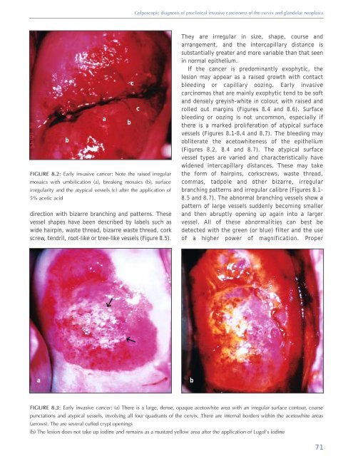
FIGURE 8.3: Early invasive cancer: (a) There is a large, dense, opaque acetowhite area with an irregular surface contour, coarse<br />
punctations <strong>and</strong> atypical vessels, involving all four quadrants <strong>of</strong> the cervix. There are internal borders within the acetowhite areas<br />
(arrows). The are several cuffed crypt openings<br />
(b) The lesion does not take up iodine <strong>and</strong> remains as a mustard yellow area after the application <strong>of</strong> Lugol’s iodine<br />
71


