

Colposcopy and Treatment of Cervical Intraepithelial Neoplasia - RHO
Colposcopy and Treatment of Cervical Intraepithelial Neoplasia - RHO
Colposcopy and Treatment of Cervical Intraepithelial Neoplasia - RHO

You also want an ePaper? Increase the reach of your titles
YUMPU automatically turns print PDFs into web optimized ePapers that Google loves.
Colposcopic assessment <strong>of</strong> cervical intraepithelial neoplasia<br />
FIGURE 7.11: Thin acetowhite lesion with geographic margins<br />
in the upper lip. Histology indicated CIN 1<br />
FIGURE 7.13: Mildly dense acetowhite lesions arising from the<br />
squamocolumnar junction in 12 <strong>and</strong> 6 o’clock position with<br />
irregular geographical margins, which on histology proved to be<br />
CIN 1 lesion<br />
↑<br />
a<br />
FIGURE 7.12: Mildly dense, thin, elongated acetowhite lesion<br />
with regular margins abutting the squamocolumnar junction.<br />
Note the fine mosaic at the distal end <strong>of</strong> the lesion. Histology<br />
indicated CIN 1<br />
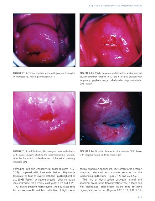
FIGURE 7.14: Note the circumorificial acetowhite CIN 1 lesion<br />
with irregular margin <strong>and</strong> fine mosaics (a)<br />
extending into the endocervical canal (Figures 7.22-<br />
7.27) compared with low-grade lesions. High-grade<br />
lesions <strong>of</strong>ten tend to involve both the lips (Burghardt et<br />
al., 1998) (Table 7.1). Severe or early malignant lesions<br />
may obliterate the external os (Figures 7.22 <strong>and</strong> 7.25).<br />
As lesions become more severe, their surfaces tend<br />
to be less smooth <strong>and</strong> less reflective <strong>of</strong> light, as in<br />
normal squamous epithelium. The surfaces can become<br />
irregular, elevated <strong>and</strong> nodular relative to the<br />
surrounding epithelium (Figures 7.20 <strong>and</strong> 7.23-7.27).<br />
The line <strong>of</strong> demarcation between normal <strong>and</strong><br />
abnormal areas in the transformation zone is sharp <strong>and</strong><br />
well delineated. High-grade lesions tend to have<br />
regular, sharper borders (Figures 7.17, 7.18, 7.19, 7.21,<br />
61


