

Colposcopy and Treatment of Cervical Intraepithelial Neoplasia - RHO
Colposcopy and Treatment of Cervical Intraepithelial Neoplasia - RHO
Colposcopy and Treatment of Cervical Intraepithelial Neoplasia - RHO

Create successful ePaper yourself
Turn your PDF publications into a flip-book with our unique Google optimized e-Paper software.
Chapter 9<br />
a<br />
a<br />
a<br />
a<br />
After application <strong>of</strong> 5% acetic acid<br />
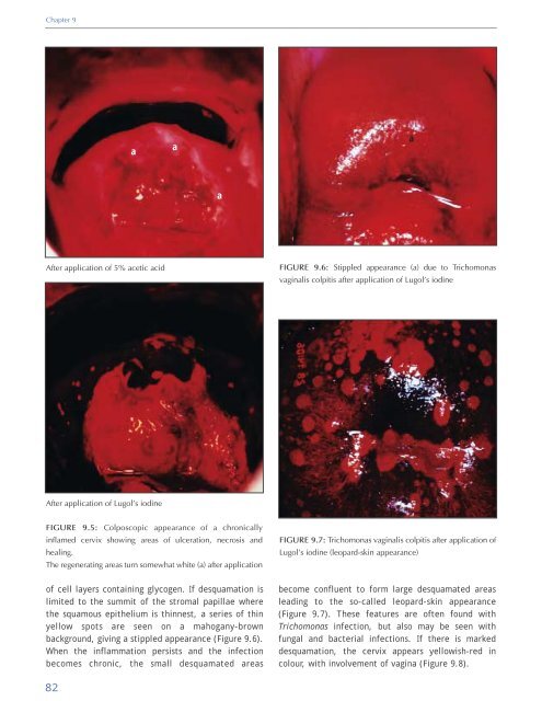
FIGURE 9.6: Stippled appearance (a) due to Trichomonas<br />
vaginalis colpitis after application <strong>of</strong> Lugol’s iodine<br />
After application <strong>of</strong> Lugol’s iodine<br />
FIGURE 9.5: Colposcopic appearance <strong>of</strong> a chronically<br />
inflamed cervix showing areas <strong>of</strong> ulceration, necrosis <strong>and</strong><br />
healing.<br />
The regenerating areas turn somewhat white (a) after application<br />
FIGURE 9.7: Trichomonas vaginalis colpitis after application <strong>of</strong><br />
Lugol’s iodine (leopard-skin appearance)<br />
<strong>of</strong> cell layers containing glycogen. If desquamation is<br />
limited to the summit <strong>of</strong> the stromal papillae where<br />
the squamous epithelium is thinnest, a series <strong>of</strong> thin<br />
yellow spots are seen on a mahogany-brown<br />
background, giving a stippled appearance (Figure 9.6).<br />
When the inflammation persists <strong>and</strong> the infection<br />
becomes chronic, the small desquamated areas<br />
become confluent to form large desquamated areas<br />
leading to the so-called leopard-skin appearance<br />
(Figure 9.7). These features are <strong>of</strong>ten found with<br />
Trichomonas infection, but also may be seen with<br />
fungal <strong>and</strong> bacterial infections. If there is marked<br />
desquamation, the cervix appears yellowish-red in<br />
colour, with involvement <strong>of</strong> vagina (Figure 9.8).<br />
82


