

Colposcopy and Treatment of Cervical Intraepithelial Neoplasia - RHO
Colposcopy and Treatment of Cervical Intraepithelial Neoplasia - RHO
Colposcopy and Treatment of Cervical Intraepithelial Neoplasia - RHO

Create successful ePaper yourself
Turn your PDF publications into a flip-book with our unique Google optimized e-Paper software.
Chapter 6<br />
ages (Figures 1.7d, 1.7e, 1.8c <strong>and</strong> 1.8d). If the junction<br />
is proximal to the os, in the canal, it requires additional<br />
effort to visualize the entire junction. Opening the<br />
blades <strong>of</strong> the vaginal speculum <strong>and</strong> using a cottontipped<br />
applicator to pry the anterior lip upward or the<br />
posterior lip downward will <strong>of</strong>ten allow visualization <strong>of</strong><br />
the squamocolumnar junction, if it is close enough to<br />
the os. The endocervical speculum (Figure 4.6) or the<br />
tips <strong>of</strong> a long dissection forceps also can be used, <strong>and</strong><br />
will <strong>of</strong>ten allow a greater length <strong>of</strong> canal to be<br />
inspected. The skill for these manoeuvres comes with<br />
practice. The vast majority <strong>of</strong> CIN lesions occur in the<br />
transformation zone <strong>and</strong> the most severe changes tend<br />
to be closer to or abutting, rather than farther from,<br />
the new squamocolumnar junction.<br />
Columnar epithelium<br />
On first looking at the normal cervix in a young woman,<br />
one sees the cervical os. It usually appears to be<br />
encircled by the columnar epithelium, appearing dark<br />
red in colour with a grape-like or sea anemone<br />
tentacles-like or a villous appearance in contrast to the<br />
smooth, light pink squamous epithelium. Each columnar<br />
villous structure contains a fine capillary <strong>and</strong> the blood<br />
in the capillary <strong>and</strong> the vascularity <strong>of</strong> the underlying<br />
connective tissue give the columnar epithelium its<br />
strikingly reddish appearance. Small polyps may be<br />
detected during examination <strong>of</strong> the endocervical canal.<br />
Vasculature<br />
The next most important feature to observe is the<br />
vasculature. The examination <strong>of</strong> the blood vessels is<br />
facilitated by applying normal saline on the cervix <strong>and</strong><br />
using the green (or blue) filter on the colposcope to<br />
enhance the contrast <strong>of</strong> the vessels. Use <strong>of</strong> a higher<br />
power <strong>of</strong> magnification (about 15x), if available in the<br />
colposcope, also is helpful. Depending on the thickness<br />
or opacity <strong>of</strong> the overlying squamous epithelium,<br />
smaller vessels may or may not be visible. The smaller<br />
vessels that may be visible are capillaries that are in the<br />
stroma below the epithelium.<br />
Two types <strong>of</strong> capillaries are apparent in the native or<br />
original squamous epithelium: reticular (network) or<br />
hairpin-shaped capillaries (Figure 6.2). The reticular<br />
pattern is especially visible because the epithelium is<br />
thinner in women taking oral contraceptives <strong>and</strong> in<br />
postmenopausal women. The hairpin capillaries<br />
actually ascend vertically, loop over, <strong>and</strong> then descend<br />
back into the stroma from where they came. Since<br />
these loops are seen ‘end on’, the colposcopic view<br />
usually is <strong>of</strong> dots with only a slight, if any, appearance<br />
<strong>of</strong> a loop at each. Inflammation <strong>of</strong> the cervix (e.g.,<br />
trichomoniasis) <strong>of</strong>ten causes hairpin vessels to form<br />
staghorn-like shapes, so that the vessels become more<br />
prominent <strong>and</strong> the loop appearance is more apparent<br />
(Figure 6.2). Often no vascular pattern is seen on the<br />
original squamous epithelium.<br />
The ectocervical vessel appearances described<br />
above are more prominent towards the outer<br />
transformation zone, nearer to the original<br />
squamocolumnar junction. In the more recently formed<br />
immature metaplastic squamous epithelium nearer the<br />
new squamocolumnar junction, other vascular patterns<br />
become more prominent. These are large (compared to<br />
capillaries) branching surface vessels with three<br />
recognizable basic patterns (Figure 6.2). The first<br />
pattern is much like a tree branching <strong>and</strong> the second is<br />
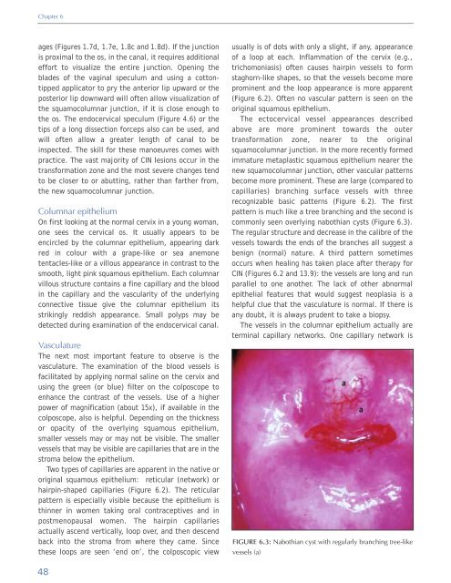
commonly seen overlying nabothian cysts (Figure 6.3).<br />
The regular structure <strong>and</strong> decrease in the calibre <strong>of</strong> the<br />
vessels towards the ends <strong>of</strong> the branches all suggest a<br />
benign (normal) nature. A third pattern sometimes<br />
occurs when healing has taken place after therapy for<br />
CIN (Figures 6.2 <strong>and</strong> 13.9): the vessels are long <strong>and</strong> run<br />
parallel to one another. The lack <strong>of</strong> other abnormal<br />
epithelial features that would suggest neoplasia is a<br />
helpful clue that the vasculature is normal. If there is<br />
any doubt, it is always prudent to take a biopsy.<br />
The vessels in the columnar epithelium actually are<br />
terminal capillary networks. One capillary network is<br />
FIGURE 6.3: Nabothian cyst with regularly branching tree-like<br />
vessels (a)<br />
a<br />
a<br />
48


