

Colposcopy and Treatment of Cervical Intraepithelial Neoplasia - RHO
Colposcopy and Treatment of Cervical Intraepithelial Neoplasia - RHO
Colposcopy and Treatment of Cervical Intraepithelial Neoplasia - RHO

Create successful ePaper yourself
Turn your PDF publications into a flip-book with our unique Google optimized e-Paper software.
An introduction to the anatomy <strong>of</strong> the uterine cervix<br />
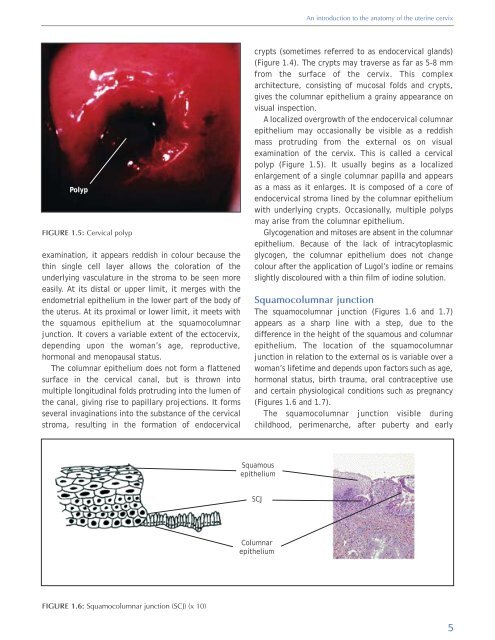
Polyp<br />
FIGURE 1.5: <strong>Cervical</strong> polyp<br />
examination, it appears reddish in colour because the<br />
thin single cell layer allows the coloration <strong>of</strong> the<br />
underlying vasculature in the stroma to be seen more<br />
easily. At its distal or upper limit, it merges with the<br />
endometrial epithelium in the lower part <strong>of</strong> the body <strong>of</strong><br />
the uterus. At its proximal or lower limit, it meets with<br />
the squamous epithelium at the squamocolumnar<br />
junction. It covers a variable extent <strong>of</strong> the ectocervix,<br />
depending upon the woman’s age, reproductive,<br />
hormonal <strong>and</strong> menopausal status.<br />
The columnar epithelium does not form a flattened<br />
surface in the cervical canal, but is thrown into<br />
multiple longitudinal folds protruding into the lumen <strong>of</strong><br />
the canal, giving rise to papillary projections. It forms<br />
several invaginations into the substance <strong>of</strong> the cervical<br />
stroma, resulting in the formation <strong>of</strong> endocervical<br />
crypts (sometimes referred to as endocervical gl<strong>and</strong>s)<br />
(Figure 1.4). The crypts may traverse as far as 5-8 mm<br />
from the surface <strong>of</strong> the cervix. This complex<br />
architecture, consisting <strong>of</strong> mucosal folds <strong>and</strong> crypts,<br />
gives the columnar epithelium a grainy appearance on<br />
visual inspection.<br />
A localized overgrowth <strong>of</strong> the endocervical columnar<br />
epithelium may occasionally be visible as a reddish<br />
mass protruding from the external os on visual<br />
examination <strong>of</strong> the cervix. This is called a cervical<br />
polyp (Figure 1.5). It usually begins as a localized<br />
enlargement <strong>of</strong> a single columnar papilla <strong>and</strong> appears<br />
as a mass as it enlarges. It is composed <strong>of</strong> a core <strong>of</strong><br />
endocervical stroma lined by the columnar epithelium<br />
with underlying crypts. Occasionally, multiple polyps<br />
may arise from the columnar epithelium.<br />
Glycogenation <strong>and</strong> mitoses are absent in the columnar<br />
epithelium. Because <strong>of</strong> the lack <strong>of</strong> intracytoplasmic<br />
glycogen, the columnar epithelium does not change<br />
colour after the application <strong>of</strong> Lugol’s iodine or remains<br />
slightly discoloured with a thin film <strong>of</strong> iodine solution.<br />
Squamocolumnar junction<br />
The squamocolumnar junction (Figures 1.6 <strong>and</strong> 1.7)<br />
appears as a sharp line with a step, due to the<br />
difference in the height <strong>of</strong> the squamous <strong>and</strong> columnar<br />
epithelium. The location <strong>of</strong> the squamocolumnar<br />
junction in relation to the external os is variable over a<br />
woman’s lifetime <strong>and</strong> depends upon factors such as age,<br />
hormonal status, birth trauma, oral contraceptive use<br />
<strong>and</strong> certain physiological conditions such as pregnancy<br />
(Figures 1.6 <strong>and</strong> 1.7).<br />
The squamocolumnar junction visible during<br />
childhood, perimenarche, after puberty <strong>and</strong> early<br />
Squamous<br />
epithelium<br />
SCJ<br />
Columnar<br />
epithelium<br />
FIGURE 1.6: Squamocolumnar junction (SCJ) (x 10)<br />
5


