

Colposcopy and Treatment of Cervical Intraepithelial Neoplasia - RHO
Colposcopy and Treatment of Cervical Intraepithelial Neoplasia - RHO
Colposcopy and Treatment of Cervical Intraepithelial Neoplasia - RHO

Create successful ePaper yourself
Turn your PDF publications into a flip-book with our unique Google optimized e-Paper software.
Chapter 7<br />
acid stain is correlated with the colour tone or intensity,<br />
the surface shine, <strong>and</strong> the duration <strong>of</strong> the effect, <strong>and</strong>,<br />
in turn, with the degree <strong>of</strong> neoplastic change in the<br />
lesion. Higher-grade lesions are more likely to turn<br />
dense white rapidly. Abnormal vascular features such as<br />
punctation, mosaicism <strong>and</strong> atypical vessels are<br />
significant only if these are seen in acetowhite areas.<br />
The acetic acid dehydrates cells <strong>and</strong> reversibly<br />
coagulates the nuclear proteins. Thus, areas <strong>of</strong><br />
increased nuclear activity <strong>and</strong> DNA content exhibit the<br />
most dramatic colour change. The most pronounced<br />
effects are observed in high-grade lesions <strong>and</strong> invasive<br />
cancer. A direct correlation exists between the<br />
intensity <strong>of</strong> the dull, white colour <strong>and</strong> the severity <strong>of</strong><br />
the lesion. Less differentiated areas are associated<br />
with an intensely opaque, dull-white appearance <strong>of</strong><br />
lesions in the transformation zone.<br />
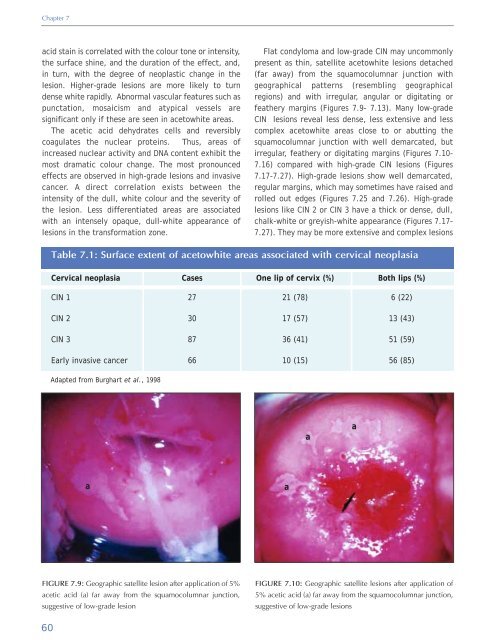
Flat condyloma <strong>and</strong> low-grade CIN may uncommonly<br />
present as thin, satellite acetowhite lesions detached<br />
(far away) from the squamocolumnar junction with<br />
geographical patterns (resembling geographical<br />
regions) <strong>and</strong> with irregular, angular or digitating or<br />
feathery margins (Figures 7.9- 7.13). Many low-grade<br />
CIN lesions reveal less dense, less extensive <strong>and</strong> less<br />
complex acetowhite areas close to or abutting the<br />
squamocolumnar junction with well demarcated, but<br />
irregular, feathery or digitating margins (Figures 7.10-<br />
7.16) compared with high-grade CIN lesions (Figures<br />
7.17-7.27). High-grade lesions show well demarcated,<br />
regular margins, which may sometimes have raised <strong>and</strong><br />
rolled out edges (Figures 7.25 <strong>and</strong> 7.26). High-grade<br />
lesions like CIN 2 or CIN 3 have a thick or dense, dull,<br />
chalk-white or greyish-white appearance (Figures 7.17-<br />
7.27). They may be more extensive <strong>and</strong> complex lesions<br />
Table 7.1: Surface extent <strong>of</strong> acetowhite areas associated with cervical neoplasia<br />
<strong>Cervical</strong> neoplasia<br />
Cases<br />
One lip <strong>of</strong> cervix (%)<br />
Both lips (%)<br />
CIN 1<br />
27<br />
21 (78)<br />
6 (22)<br />
CIN 2<br />
30<br />
17 (57)<br />
13 (43)<br />
CIN 3<br />
87<br />
36 (41)<br />
51 (59)<br />
Early invasive cancer<br />
66<br />
10 (15)<br />
56 (85)<br />
Adapted from Burghart et al., 1998<br />
a<br />
a<br />
a<br />
a<br />
FIGURE 7.9: Geographic satellite lesion after application <strong>of</strong> 5%<br />
acetic acid (a) far away from the squamocolumnar junction,<br />
suggestive <strong>of</strong> low-grade lesion<br />
FIGURE 7.10: Geographic satellite lesions after application <strong>of</strong><br />
5% acetic acid (a) far away from the squamocolumnar junction,<br />
suggestive <strong>of</strong> low-grade lesions<br />
60


