

Colposcopy and Treatment of Cervical Intraepithelial Neoplasia - RHO
Colposcopy and Treatment of Cervical Intraepithelial Neoplasia - RHO
Colposcopy and Treatment of Cervical Intraepithelial Neoplasia - RHO

You also want an ePaper? Increase the reach of your titles
YUMPU automatically turns print PDFs into web optimized ePapers that Google loves.
Chapter 7<br />
The colposcopic diagnosis <strong>of</strong> cervical neoplasia requires<br />
an underst<strong>and</strong>ing <strong>and</strong> recognition <strong>of</strong> four main<br />
features: colour tone <strong>and</strong> intensity <strong>of</strong> acetowhitening,<br />
margins <strong>and</strong> surface contour <strong>of</strong> acetowhite areas,<br />
vascular pattern <strong>and</strong> iodine staining. <strong>Colposcopy</strong> with<br />
directed biopsy is described as the reference<br />
investigation or ‘gold st<strong>and</strong>ard’ for the diagnosis <strong>of</strong><br />
cervical precancer (Singer & Monaghan, 2000).<br />
<strong>Colposcopy</strong> has a reported sensitivity ranging from 87%<br />
to 99% to diagnose cervical neoplasia, but its specificity<br />
is lower, between 23% <strong>and</strong> 87% (Mitchell et al., 1998;<br />
Belinson et al., 2001).<br />
The colposcopic features <strong>of</strong> cervical intraepithelial<br />
neoplasia (CIN) are described in this chapter to equip<br />
the student with the skills to distinguish the<br />
colposcopic findings associated with high-grade CIN<br />
(CIN 2-3) from those <strong>of</strong> low-grade lesions (CIN 1).<br />
Although the appearance <strong>of</strong> a single abnormal feature<br />
alone is not a strong indicator that a lesion is present,<br />
the occurrence <strong>of</strong> abnormal features together in a<br />
localized area in the transformation zone increases the<br />
probability <strong>of</strong> a lesion. It will become obvious during<br />
colposcopic practice that considerable skills are<br />
required to differentiate between low-grade lesions,<br />
immature squamous metaplasia <strong>and</strong> certain<br />
inflammatory conditions. The student is encouraged to<br />
obtain biopsies whenever in doubt, <strong>and</strong> to review the<br />
histopathological findings with the pathologist. Close<br />
collaboration with pathologists is obligatory <strong>and</strong> useful<br />
in improving one’s diagnostic skills. At the end <strong>of</strong> this<br />
chapter, a system that enables the colposcopist to<br />
score abnormalities is presented. This system is useful<br />
as a basis for the choice <strong>of</strong> which area(s) to select for<br />
biopsy. It is essential to biopsy the ‘worst’ area(s) - that<br />
is, the area(s) with the most severe changes in<br />
features.<br />
The colposcopic findings <strong>of</strong> an abnormal or atypical<br />
transformation zone can involve the whole<br />
transformation zone but more commonly affect only a<br />
portion <strong>of</strong> it <strong>and</strong> there may be multiple distinct lesions.<br />
There is usually a distinct demarcation between normal<br />
<strong>and</strong> abnormal epithelium.<br />
The colposcopic features that differentiate an<br />
abnormal transformation zone from the normal include<br />
the following: colour tone <strong>of</strong> acetowhite areas; surface<br />
pattern <strong>of</strong> acetowhite areas; borderline between<br />
acetowhite areas <strong>and</strong> the rest <strong>of</strong> the epithelium;<br />
vascular features <strong>and</strong> colour changes after application<br />
<strong>of</strong> iodine.<br />
After application <strong>of</strong> normal saline solution<br />
Following application <strong>of</strong> saline, abnormal epithelium<br />
may appear much darker than the normal epithelium.<br />
Vasculature<br />
Using the green (or blue) filter <strong>and</strong> higher-power<br />
magnification when necessary, the best opportunity to<br />
evaluate any abnormal vasculature patterns is before<br />
the application <strong>of</strong> acetic acid, the effect <strong>of</strong> which may<br />
obscure some or all <strong>of</strong> the changes, especially in an<br />
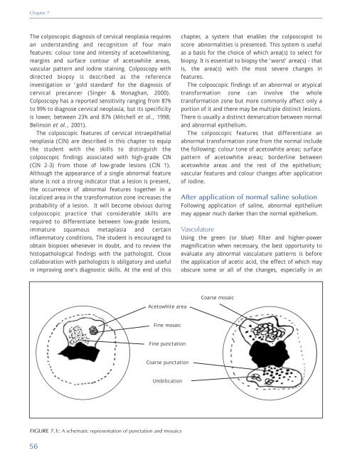
Acetowhite area<br />
Coarse mosaic<br />
Fine mosaic<br />
Fine punctation<br />
Coarse punctation<br />
Umbilication<br />
FIGURE 7.1: A schematic representation <strong>of</strong> punctation <strong>and</strong> mosaics<br />
56


