

Colposcopy and Treatment of Cervical Intraepithelial Neoplasia - RHO
Colposcopy and Treatment of Cervical Intraepithelial Neoplasia - RHO
Colposcopy and Treatment of Cervical Intraepithelial Neoplasia - RHO

Create successful ePaper yourself
Turn your PDF publications into a flip-book with our unique Google optimized e-Paper software.
Chapter 2<br />
Moscicki et al., 2001; Woodman et al., 2001; Schlecht<br />
et al., 2002).<br />
HPV infection is transmitted through sexual contact<br />
<strong>and</strong> the risk factors therefore are closely related to<br />
sexual behaviour (e.g., lifetime number <strong>of</strong> sexual<br />
partners, sexual intercourse at an early age). In most<br />
women, HPV infections are transient. The natural<br />
history <strong>of</strong> HPV infection has been extensively reviewed.<br />
Although the prevalence <strong>of</strong> HPV infection varies in<br />
different regions <strong>of</strong> the world, it generally reaches a<br />
peak <strong>of</strong> about 20-30% among women aged 20-24 years,<br />
with a subsequent decline to approximately 3-10%<br />
among women aged over 30 (Herrero et al., 1997a;<br />
Herrero et al., 1997b; Sellors et al., 2000). About 80%<br />
<strong>of</strong> young women who become infected with HPV have<br />
transient infections that clear up within 12-18 months<br />
(Ho et al., 1998; Franco et al., 1999; Thomas et al.,<br />
2000; Liaw et al., 2001).<br />
HPV infection is believed to start in the basal cells or<br />
parabasal cells <strong>of</strong> the metaplastic epithelium. If the<br />
infection persists, integration <strong>of</strong> viral genome into the<br />
host cellular genome may occur. The normal<br />
differentiation <strong>and</strong> maturation <strong>of</strong> the immature<br />
squamous metaplastic into the mature squamous<br />
metaplastic epithelium may be disrupted as a result <strong>of</strong><br />
expression <strong>of</strong> E6/E7 oncoproteins <strong>and</strong> the loss <strong>of</strong><br />
normal growth control. This may then lead to<br />
development <strong>of</strong> abnormal dysplastic epithelium. If the<br />
neoplastic process continues uninterrupted, the early<br />
low-grade lesions may eventually involve the full<br />
thickness <strong>of</strong> the epithelium. Subsequently the disease<br />
may traverse the basement membrane <strong>and</strong> become<br />
invasive cancer, extending to surrounding tissues <strong>and</strong><br />
organs. The invasion may then affect blood <strong>and</strong><br />
lymphatic vessels <strong>and</strong> the disease may spread to the<br />
lymph nodes <strong>and</strong> distant organs.<br />
Natural history <strong>of</strong> cervical cancer<br />
precursors<br />
Despite women’s frequent exposure to HPV,<br />
development <strong>of</strong> cervical neoplasia is uncommon. Most<br />
cervical abnormalities caused by HPV infection are<br />
unlikely to progress to high-grade CIN or cervical cancer,<br />
as most <strong>of</strong> them regress by themselves. The long time<br />
frame between initial infection <strong>and</strong> overt disease<br />
indicates that several c<strong>of</strong>actors (e.g., genetic<br />
differences, hormonal effects, micronutrient<br />
deficiencies, smoking, or chronic inflammation) may be<br />
necessary for disease progression. Spontaneous<br />
regression <strong>of</strong> CIN may also indicate that many women<br />
may not be exposed to these c<strong>of</strong>actors.<br />
Several studies have addressed the natural history <strong>of</strong><br />
CIN, with particular emphasis on disease regression,<br />
persistence <strong>and</strong> progression (McIndoe et al., 1984;<br />
Östor et al., 1993; Mitchell et al., 1994; Melinkow et<br />
al., 1998; Holowaty et al., 1999). They have revealed<br />
that most low-grade lesions are transient; most <strong>of</strong> them<br />
regress to normal within relatively short periods or do<br />
not progress to more severe forms. High-grade CIN, on<br />
the other h<strong>and</strong>, carries a much higher probability <strong>of</strong><br />
progressing to invasive cancer, although a proportion <strong>of</strong><br />
such lesions also regress or persist. It is appears that<br />
the mean interval for progression <strong>of</strong> cervical precursors<br />
to invasive cancer is some 10 to 20 years.<br />
A few studies have attempted to summarize the<br />
rates <strong>of</strong> regression, persistence <strong>and</strong> progression <strong>of</strong> CIN.<br />
Even though these studies have many limitations, they<br />
provide interesting insight into the biological behaviour<br />
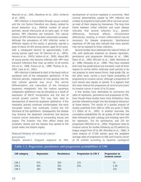
<strong>of</strong> these lesions. The results <strong>of</strong> a pooled analysis <strong>of</strong><br />
studies published from 1950 to 1993 are given in Table<br />
2.3 (Östor et al., 1993). In another overview, the<br />
cumulative probabilities for all grades <strong>of</strong> CIN that had<br />
been followed by both cytology <strong>and</strong> histology were 45%<br />
for regression, 31% for persistence, <strong>and</strong> 23% for<br />
progression (Mitchell et al., 1994). Progression rates to<br />
invasive cancer for studies following up CIS patients by<br />
biopsy ranged from 29 to 36% (McIndoe et al., 1984). A<br />
meta analysis <strong>of</strong> 27 000 women gave the weighted<br />
average rates <strong>of</strong> progression to HSIL <strong>and</strong> invasive cancer<br />
at 24 months according to baseline cytological<br />
Table 2.3: Regression, persistence <strong>and</strong> progression probabilities <strong>of</strong> CIN<br />
CIN category<br />
Regression<br />
Persistence<br />
Progression to CIN 3<br />
Progression to<br />
invasive cancer<br />
CIN 1<br />
CIN 2<br />
CIN 3<br />
57%<br />
43%<br />
32%<br />
32%<br />
35%<br />
56%<br />
11%<br />
22%<br />
-<br />
1%<br />
1.5%<br />
12%<br />
18


