Early Cretaceous Freshwater Fish Fauna in Kyushu, Japan
Early Cretaceous Freshwater Fish Fauna in Kyushu, Japan
Early Cretaceous Freshwater Fish Fauna in Kyushu, Japan

Create successful ePaper yourself
Turn your PDF publications into a flip-book with our unique Google optimized e-Paper software.
<strong>Early</strong> <strong>Cretaceous</strong> <strong>Freshwater</strong> <strong>Fish</strong> <strong>Fauna</strong> <strong>in</strong> <strong>Kyushu</strong>, <strong>Japan</strong> 211<br />
is slightly convex. The restored outl<strong>in</strong>e ofthe abdomen is remarkably convex (Figs.<br />
73 and 74). The dorsal f<strong>in</strong> is situated at about the middle of the body. The anal<br />
orig<strong>in</strong> is well beh<strong>in</strong>d the posterior end of the dorsal f<strong>in</strong> base. The first dorsal f<strong>in</strong><br />
pterygiophore is above the9th abdom<strong>in</strong>al vertebra. The dorsal f<strong>in</strong> base is short and<br />
is conta<strong>in</strong>ed 2.1 times <strong>in</strong> the anal f<strong>in</strong> base. The pectoral f<strong>in</strong> is short and does not reach<br />
the pelvic <strong>in</strong>sertion (Figs. 73 and 74). Thenumber ofpr<strong>in</strong>cipal dorsal f<strong>in</strong> rays is 10.<br />
There are 10 dorsal f<strong>in</strong> pterygiophores. The number of pr<strong>in</strong>cipal anal f<strong>in</strong> rays is 24.<br />
There are 24 anal f<strong>in</strong> pterygiophores. The pr<strong>in</strong>cipal dorsal and anal f<strong>in</strong> rays are<br />
preceded by two small unbranched accessory rays respectively. Six pelvic f<strong>in</strong> rays<br />
are visible. There are 18 pr<strong>in</strong>cipal caudal f<strong>in</strong> rays (1,8,8,1). The pelvic f<strong>in</strong> is<br />
situated at about the middle of the body and the <strong>in</strong>sertion is situated slightly before<br />
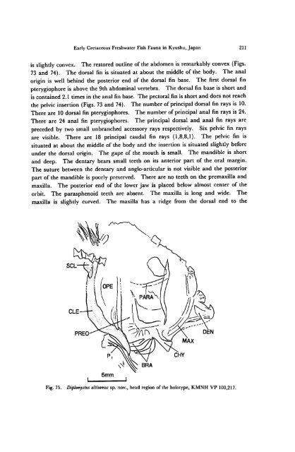
under the dorsal orig<strong>in</strong>. The gape of the mouth is small. The mandible is short<br />
and deep. The dentary bears small teeth on its anterior part of the oral marg<strong>in</strong>.<br />
The suture between the dentary and anglo-articular is not visible and the posterior<br />
part of the mandible is poorly preserved. There are no teeth on the premaxilla and<br />
maxilla. The posterior end of the lower jaw is placed below almost center of the<br />
orbit. The parasphenoid teeth are absent. The maxilla is long and wide. The<br />
maxilla is slightly curved. The maxilla has a ridge from the dorsal end to the<br />
SCL<br />
CLE<br />
PREO<br />
5mm<br />
Fig. 75. Diplomystus altisomus sp. nov., head region of the holotype, KMNH VP 100,217.