Clinical work involves development <strong>of</strong> sports vision as a <strong>research</strong> theme and to see how binocular vision processescan be optimised to improve sports performance as well as reading and writing ability. This work is conducted withDr McDowell and Mr Gilsenan (private practice) and resulted in a television interviewed on 9th January 2008. Theimprovement <strong>of</strong> reading writing and sporting ability addresses national and international priorities in education andlearning.Pr<strong>of</strong>essor Pierscionek utilises her legal qualifications to develop <strong>research</strong> in healthcare ethics and medical law asapplied to eye care practice. This has produced the first book <strong>of</strong> its kind on healthcare ethics and law for eye carepractice and a peer-review paper which from its publication in April to July 2008 had been accessed over 1465 timesand remains one <strong>of</strong> the top 10 most viewed articles in BMC Medical Ethics.Pr<strong>of</strong>essor Pierscionek was invited to give a plenary lecture: “The paradox <strong>of</strong> the eye lens”, SPIE Student ChapterWroclaw, Poland (May 2008)During the year Pr<strong>of</strong>essor Pierscionek also secured a DEL-funded studentship for “Identification <strong>of</strong> Iris Biometrics”(circa £48,000).Publications:Asejczyk-Widlicka M and Pierscionek BK; Fluctuations in intraocular pressure and the potential effect on aberrations<strong>of</strong> the eye; British Journal <strong>of</strong> Ophthalmology, 91: 1054-1058, 2007Belaidi A and Pierscionek BK; Modelling internal stress distributions in the human lens: can opponent theoriescoexist? Journal <strong>of</strong> Vision, 7: 1-12, 2007Schachar RA, Kamangar F and Pierscionek BK; Edinger-Westphal and pharmacologically stimulated accommodativerefractive changes and lens ciliary process movements in rhesus monkeys; by L.A. Ostrin and A. Glasser; ExperimentalEye <strong>Research</strong>, 85: 298-301, 2007Keenan J, Orr DF and Pierscionek BK; Patterns <strong>of</strong> protein distribution in porcine eye lenses; Molecular Vision, 14: 1245-1253, 2008Pierscionek BK; What is presumed when we presume consent? BMC Medical Ethics, 9:8 doi:10.1186/1472-6939-9-8,2008Schachar RA, Chen W, Woo BK, Pierscionek BK, Zhang X and Ma L; Diffusion <strong>of</strong> nanoparticles into the capsule andcortex <strong>of</strong> the crystalline lens; Nanotechnology, 19: 1-4, 2008Schachar RA, Kamangar F and Pierscionek BK; Changes in lens dimensions and refractive index with age andaccommodation; Optometry and Vision Science, 85: 281-282, 2008Siedlecki D, Kasprzak H and Pierscionek BK; Radial gradient index intraocular lens: a theoretical model; Journal <strong>of</strong>Modern Optics, 55: 639-647, 2008Book:Pierscionek BK; Law and ethics for the eye-care pr<strong>of</strong>essional; Elsevier (2008)110
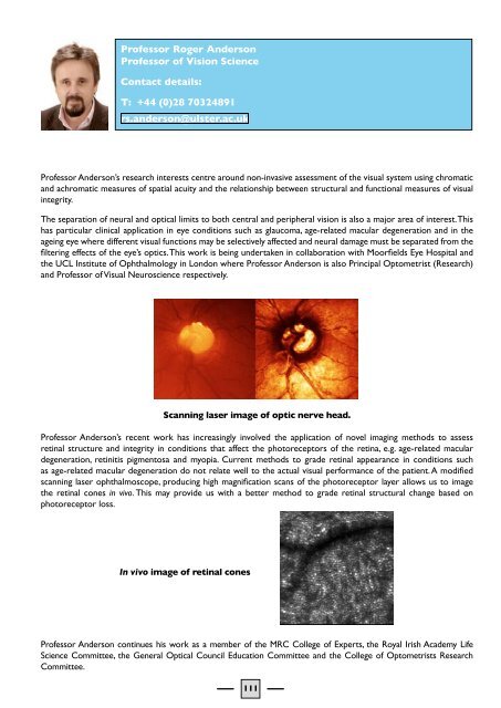
Pr<strong>of</strong>essor Roger AndersonPr<strong>of</strong>essor <strong>of</strong> Vision ScienceContact details:T: +44 (0)28 70324891rs.anderson@ulster.ac.ukPr<strong>of</strong>essor Anderson’s <strong>research</strong> interests centre around non-invasive assessment <strong>of</strong> the visual system using chromaticand achromatic measures <strong>of</strong> spatial acuity and the relationship between structural and functional measures <strong>of</strong> visualintegrity.The separation <strong>of</strong> neural and optical limits to both central and peripheral vision is also a major area <strong>of</strong> interest. Thishas particular clinical application in eye conditions such as glaucoma, age-related macular degeneration and in theageing eye where different visual functions may be selectively affected and neural damage must be separated from thefiltering effects <strong>of</strong> the eye’s optics. This work is being undertaken in collaboration with Moorfields Eye Hospital andthe UCL Institute <strong>of</strong> Ophthalmology in London where Pr<strong>of</strong>essor Anderson is also Principal Optometrist (<strong>Research</strong>)and Pr<strong>of</strong>essor <strong>of</strong> Visual Neuroscience respectively.Scanning laser image <strong>of</strong> optic nerve head.Pr<strong>of</strong>essor Anderson’s recent work has increasingly involved the application <strong>of</strong> novel imaging methods to assessretinal structure and integrity in conditions that affect the photoreceptors <strong>of</strong> the retina, e.g. age-related maculardegeneration, retinitis pigmentosa and myopia. Current methods to grade retinal appearance in conditions suchas age-related macular degeneration do not relate well to the actual visual performance <strong>of</strong> the patient. A modifiedscanning laser ophthalmoscope, producing high magnification scans <strong>of</strong> the photoreceptor layer allows us to imagethe retinal cones in vivo. This may provide us with a better method to grade retinal structural change based onphotoreceptor loss.In vivo image <strong>of</strong> retinal conesPr<strong>of</strong>essor Anderson continues his work as a member <strong>of</strong> the MRC College <strong>of</strong> Experts, the Royal Irish Academy LifeScience Committee, the General Optical Council Education Committee and the College <strong>of</strong> Optometrists <strong>Research</strong>Committee.111
- Page 1:
BIOMEDICAL SCIENCESRESEARCH INSTITU
- Page 4 and 5:
1 Foreword by the Pro Vice-Chancell
- Page 6 and 7:
2 Foreword by the Research Institut
- Page 8 and 9:
The BMSRI Research StructureThe BMS
- Page 10 and 11:
BMSRI Core FacilitiesContact: Karen
- Page 12 and 13:
of Metabolomics, pharmacy, nutritio
- Page 14 and 15:
BMSRI Academic Heads new Regional N
- Page 16 and 17:
4. BIOMEDICAL GENOMICS RESEARCH GRO
- Page 18 and 19:
Recent Funding Initiatives:C-TRIC:
- Page 20 and 21:
Dr Mateus Webba da SilvaLecturer in
- Page 22 and 23:
5. BIOIMAGING RESEARCH GROUPResearc
- Page 24 and 25:
developmental alterations that mani
- Page 26 and 27:
Publications:Bigot S, Lucas L, Morr
- Page 28 and 29:
Publications:Barnes CA, O’Hagan B
- Page 30 and 31:
We also measure the genotoxic effec
- Page 32 and 33:
6. CANCER AND AGEING RESEARCH GROUP
- Page 34 and 35:
Professor Anthony P McHaleProfessor
- Page 36 and 37:
Professor Stephanie McKeownProfesso
- Page 38 and 39:
an Alzheimer Research Trust collabo
- Page 40 and 41:
JM, Waugh DJJ; Dexamethasone potent
- Page 42 and 43:
have wider applications in vivo, in
- Page 44 and 45:
Inter-relationships between diet an
- Page 46 and 47:
Flatt PR; Effective surgical treatm
- Page 48 and 49:
These areas are the subject of seve
- Page 50 and 51:
Dr YHA Abdel-WahabSenior Lecturer i
- Page 52 and 53:
Dr VA GaultLecturer in Molecular Bi
- Page 54 and 55:
McClean PL, Irwin N, Hunter K, Gaul
- Page 56 and 57:
Publications:Duffy NA, Green BD, Ir
- Page 58 and 59:
8. MICROBIOLOGY AND BIOTECHNOLOGYRE
- Page 60 and 61:
Publications:Graham RJL, Graham C,
- Page 62 and 63: analyses. However there is still a
- Page 64 and 65: Plessas S, Bekatorou A, Koutinas AA
- Page 66 and 67: Biochemical studies/ viral evasion
- Page 68 and 69: Dr Stephen McCleanLecturer in Prote
- Page 70 and 71: environmental remediation and as ro
- Page 72 and 73: 9. NORTHERN IRELAND CENTRE FOR FOOD
- Page 74 and 75: 26-28 Sept 2007: International Seaf
- Page 76 and 77: Dr Barnes has developed expertise i
- Page 78 and 79: Dr Alison GallagherSenior Lecturer
- Page 80 and 81: In addition, results of a pilot stu
- Page 82 and 83: Dr Maeve KerrResearch AssociateCont
- Page 84 and 85: Memberships of External Committees/
- Page 86 and 87: Indicators of Esteem:Professor McNu
- Page 88 and 89: were examined. The intervention and
- Page 90 and 91: Within this work both short-term an
- Page 92 and 93: micronutrient supplementation at a
- Page 94 and 95: 10. STEM CELL & EPIGENETICS RESEARC
- Page 96 and 97: Publications:Lees-Murdock DJ, Lau H
- Page 98 and 99: Publications:Lees-Murdock DJ, Lau H
- Page 100 and 101: 11. SYSTEMS BIOLOGY RESEARCH GROUPT
- Page 102 and 103: Kravtsov V, Swain M, Schuster A, Du
- Page 104 and 105: Dr Daniel BerrarLecturer in Biomedi
- Page 106 and 107: Zhang et al; Incorporating Feature
- Page 108 and 109: Bala P, Baldridge K, Benfenati E, C
- Page 110 and 111: In addition there is a growing them
- Page 114 and 115: In 2009 he was appointed Chairman o
- Page 116 and 117: Stevenson TR, Goodall EA and Moore
- Page 118 and 119: Dr Raymond BeirneLecturer in Optome
- Page 120 and 121: Dr Julie McClellandLecturer in Opto
- Page 122 and 123: Graham JE, Moore JE, Moore JE, McCl
- Page 124 and 125: Research Staff:Dr David OrrSenior R
- Page 126 and 127: Dr Victoria McGilliganResearch Asso
- Page 128 and 129: 13. Externally Funded Projects duri
- Page 130 and 131: Grant Holder Anderson, Prof RSFundi
- Page 132 and 133: Funding Body Royal Irish AcademyAmo
- Page 134 and 135: 14. BIOMEDICAL SCIENCES RESEARCH IN
- Page 136 and 137: Student: Simon GenglerTitle: Effect
- Page 138 and 139: Student: Anisha MazumdarTitle: Anal
- Page 140 and 141: Student: Clare RyanTitle: How does
- Page 142 and 143: CONGRATULATIONS TO THE FOLLOWING PO