

The Origin and Evolution of Mammals - Moodle
The Origin and Evolution of Mammals - Moodle
The Origin and Evolution of Mammals - Moodle

Create successful ePaper yourself
Turn your PDF publications into a flip-book with our unique Google optimized e-Paper software.
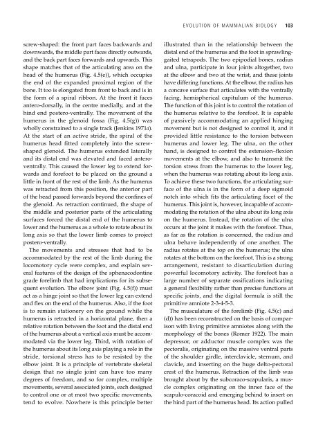
screw-shaped: the front part faces backwards <strong>and</strong><br />
downwards, the middle part faces directly outwards,<br />
<strong>and</strong> the back part faces forwards <strong>and</strong> upwards. This<br />
shape matches that <strong>of</strong> the articulating area on the<br />
head <strong>of</strong> the humerus (Fig. 4.5(e)), which occupies<br />
the end <strong>of</strong> the exp<strong>and</strong>ed proximal region <strong>of</strong> the<br />
bone. It too is elongated from front to back <strong>and</strong> is in<br />
the form <strong>of</strong> a spiral ribbon. At the front it faces<br />
antero-dorsally, in the centre medially, <strong>and</strong> at the<br />
hind end postero-ventrally. <strong>The</strong> movement <strong>of</strong> the<br />
humerus in the glenoid fossa (Fig. 4.5(g)) was<br />
wholly constrained to a single track (Jenkins 1971a).<br />
At the start <strong>of</strong> an active stride, the spiral <strong>of</strong> the<br />
humerus head fitted completely into the screwshaped<br />
glenoid. <strong>The</strong> humerus extended laterally<br />
<strong>and</strong> its distal end was elevated <strong>and</strong> faced anteroventrally.<br />
This caused the lower leg to extend forwards<br />
<strong>and</strong> forefoot to be placed on the ground a<br />
little in front <strong>of</strong> the rest <strong>of</strong> the limb. As the humerus<br />
was retracted from this position, the anterior part<br />
<strong>of</strong> the head passed forwards beyond the confines <strong>of</strong><br />
the glenoid. As retraction continued, the shape <strong>of</strong><br />
the middle <strong>and</strong> posterior parts <strong>of</strong> the articulating<br />
surfaces forced the distal end <strong>of</strong> the humerus to<br />
lower <strong>and</strong> the humerus as a whole to rotate about its<br />
long axis so that the lower limb comes to project<br />
postero-ventrally.<br />
<strong>The</strong> movements <strong>and</strong> stresses that had to be<br />
accommodated by the rest <strong>of</strong> the limb during the<br />
locomotory cycle were complex, <strong>and</strong> explain several<br />
features <strong>of</strong> the design <strong>of</strong> the sphenacodontine<br />
grade forelimb that had implications for its subsequent<br />
evolution. <strong>The</strong> elbow joint (Fig. 4.5(f)) must<br />
act as a hinge joint so that the lower leg can extend<br />
<strong>and</strong> flex on the end <strong>of</strong> the humerus. Also, if the foot<br />
is to remain stationery on the ground while the<br />
humerus is retracted in a horizontal plane, then a<br />
relative rotation between the foot <strong>and</strong> the distal end<br />
<strong>of</strong> the humerus about a vertical axis must be accommodated<br />
via the lower leg. Third, with rotation <strong>of</strong><br />
the humerus about its long axis playing a role in the<br />
stride, torsional stress has to be resisted by the<br />
elbow joint. It is a principle <strong>of</strong> vertebrate skeletal<br />
design that no single joint can have too many<br />
degrees <strong>of</strong> freedom, <strong>and</strong> so for complex, multiple<br />
movements, several associated joints, each designed<br />
to control one or at most two specific movements,<br />
tend to evolve. Nowhere is this principle better<br />
EVOLUTION OF MAMMALIAN BIOLOGY 103<br />
illustrated than in the relationship between the<br />
distal end <strong>of</strong> the humerus <strong>and</strong> the foot in sprawlinggaited<br />
tetrapods. <strong>The</strong> two epipodial bones, radius<br />
<strong>and</strong> ulna, participate in four joints altogether, two<br />
at the elbow <strong>and</strong> two at the wrist, <strong>and</strong> these joints<br />
have differing functions. At the elbow, the radius has<br />
a concave surface that articulates with the ventrally<br />
facing, hemispherical capitulum <strong>of</strong> the humerus.<br />
<strong>The</strong> function <strong>of</strong> this joint is to control the rotation <strong>of</strong><br />
the humerus relative to the forefoot. It is capable<br />
<strong>of</strong> passively accommodating an applied hinging<br />
movement but is not designed to control it, <strong>and</strong> it<br />
provided little resistance to the torsion between<br />
humerus <strong>and</strong> lower leg. <strong>The</strong> ulna, on the other<br />
h<strong>and</strong>, is designed to control the extension–flexion<br />
movements at the elbow, <strong>and</strong> also to transmit the<br />
torsion stress from the humerus to the lower leg,<br />
when the humerus was rotating about its long axis.<br />
To achieve these two functions, the articulating surface<br />
<strong>of</strong> the ulna is in the form <strong>of</strong> a deep sigmoid<br />
notch into which fits the articulating facet <strong>of</strong> the<br />
humerus. This joint is, however, incapable <strong>of</strong> accommodating<br />
the rotation <strong>of</strong> the ulna about its long axis<br />
on the humerus. Instead, the rotation <strong>of</strong> the ulna<br />
occurs at the joint it makes with the forefoot. Thus,<br />
as far as the rotation is concerned, the radius <strong>and</strong><br />
ulna behave independently <strong>of</strong> one another. <strong>The</strong><br />
radius rotates at the top on the humerus; the ulna<br />
rotates at the bottom on the forefoot. This is a strong<br />
arrangement, resistant to disarticulation during<br />
powerful locomotory activity. <strong>The</strong> forefoot has a<br />
large number <strong>of</strong> separate ossifications indicating<br />
a general flexibility rather than precise functions at<br />
specific joints, <strong>and</strong> the digital formula is still the<br />
primitive amniote 2-3-4-5-3.<br />
<strong>The</strong> musculature <strong>of</strong> the forelimb (Fig. 4.5(c) <strong>and</strong><br />
(d)) has been reconstructed on the basis <strong>of</strong> comparison<br />
with living primitive amniotes along with the<br />
morphology <strong>of</strong> the bones (Romer 1922). <strong>The</strong> main<br />
depressor, or adductor muscle complex was the<br />
pectoralis, originating on the massive ventral parts<br />
<strong>of</strong> the shoulder girdle, interclavicle, sternum, <strong>and</strong><br />
clavicle, <strong>and</strong> inserting on the huge delto-pectoral<br />
crest <strong>of</strong> the humerus. Retraction <strong>of</strong> the limb was<br />
brought about by the subcoraco-scapularis, a muscle<br />
complex originating on the inner face <strong>of</strong> the<br />
scapulo-coracoid <strong>and</strong> emerging behind to insert on<br />
the hind part <strong>of</strong> the humerus head. Its action pulled