

The Origin and Evolution of Mammals - Moodle
The Origin and Evolution of Mammals - Moodle
The Origin and Evolution of Mammals - Moodle

You also want an ePaper? Increase the reach of your titles
YUMPU automatically turns print PDFs into web optimized ePapers that Google loves.
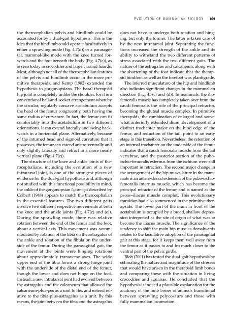
the therocephalian pelvis <strong>and</strong> hindlimb could be<br />
accounted for by a dual-gait hypothesis. This is the<br />
idea that the hindlimb could operate facultatively in<br />
either a sprawling mode (Fig. 4.7(d)) or a parasagittal,<br />
mammal-like mode with the knee turned forwards<br />
<strong>and</strong> the foot beneath the body (Fig. 4.7(c)), as<br />
is seen today in crocodiles <strong>and</strong> large varanid lizards.<br />
Most, although not all <strong>of</strong> the therocephalian features<br />
<strong>of</strong> the pelvis <strong>and</strong> hindlimb occur in the more primitive<br />
therapsids, <strong>and</strong> Kemp (1982) extended the<br />
hypothesis to gorgonopsians. <strong>The</strong> basal therapsid<br />
hip joint is completely unlike the shoulder, for it is a<br />
conventional ball-<strong>and</strong>-socket arrangement whereby<br />
the circular, regularly concave acetabulum accepts<br />
the head <strong>of</strong> the femur congruently, both having the<br />
same radius <strong>of</strong> curvature. In fact, the femur can fit<br />
comfortably into the acetabulum in two different<br />
orientations. It can extend laterally <strong>and</strong> swing backwards<br />
in a horizontal plane. Alternatively, because<br />
<strong>of</strong> the inturned head <strong>and</strong> sigmoid curvature that it<br />
possesses, the femur can extend antero-ventrally <strong>and</strong><br />
only slightly laterally <strong>and</strong> retract in a more nearly<br />
vertical plane (Fig. 4.7(c)).<br />
<strong>The</strong> structure <strong>of</strong> the knee <strong>and</strong> ankle joints <strong>of</strong> therocephalians,<br />
including the evolution <strong>of</strong> a new<br />
intratarsal joint, is one <strong>of</strong> the strongest pieces <strong>of</strong><br />
evidence for the dual-gait hypothesis <strong>and</strong>, although<br />
not studied with this functional possibility in mind,<br />
the ankle <strong>of</strong> the gorgonopsian Lycaenops described by<br />
Colbert (1948) appears to match the therocephalian<br />
in the essential features. <strong>The</strong> two different gaits<br />
involve two different respective movements at both<br />
the knee <strong>and</strong> the ankle joints (Fig. 4.7(c) <strong>and</strong> (e)).<br />
During the sprawling mode, there was relative<br />
rotation between the end <strong>of</strong> the femur <strong>and</strong> the foot,<br />
about a vertical axis. This movement was accommodated<br />
by rotation <strong>of</strong> the tibia on the astragalus <strong>of</strong><br />
the ankle <strong>and</strong> rotation <strong>of</strong> the fibula on the underside<br />
<strong>of</strong> the femur. During the parasagittal gait, the<br />
movement at the joints were hinging rotations<br />
about approximately transverse axes. <strong>The</strong> wide<br />
upper end <strong>of</strong> the tibia forms a strong hinge joint<br />
with the underside <strong>of</strong> the distal end <strong>of</strong> the femur,<br />
though the lower end does not hinge on the foot.<br />
Instead, a new intratarsal joint had evolved between<br />
the astragalus <strong>and</strong> the calcaneum that allowed the<br />
calcaneum-plus-pes as a unit to flex <strong>and</strong> extend relative<br />
to the tibia-plus-astragalus as a unit. By this<br />
means, the joint between the tibia <strong>and</strong> the astragalus<br />
EVOLUTION OF MAMMALIAN BIOLOGY 109<br />
does not have to undergo both rotation <strong>and</strong> hinging,<br />
but only the former. <strong>The</strong> latter is taken care <strong>of</strong><br />
by the new intratarsal joint. Separating the functions<br />
increased the strength <strong>of</strong> the ankle <strong>and</strong> its<br />
ability to withst<strong>and</strong> the two different patterns <strong>of</strong><br />
stress associated with the two different gaits. <strong>The</strong><br />
nature <strong>of</strong> the astragalus <strong>and</strong> calcaneum, along with<br />
the shortening <strong>of</strong> the foot indicate that the therapsid<br />
hindfoot as well as the forefoot was plantigrade.<br />
<strong>The</strong> inferred musculature <strong>of</strong> the hip <strong>and</strong> hindlimb<br />
also indicates significant changes in the mammalian<br />
direction (Fig. 4.7(c) <strong>and</strong> (d)). In mammals, the ili<strong>of</strong>emoralis<br />
muscle has completely taken over from the<br />
caudi femoralis the role <strong>of</strong> the principal retractor,<br />
becoming the gluteal muscle complex. In primitive<br />
therapsids, the combination <strong>of</strong> enlarged <strong>and</strong> somewhat<br />
anteriorly extended ilium, development <strong>of</strong> a<br />
distinct trochanter major on the hind edge <strong>of</strong> the<br />
femur, <strong>and</strong> reduction <strong>of</strong> the tail, point to an early<br />
stage in this transition. Nevertheless, the retention <strong>of</strong><br />
an internal trochanter on the underside <strong>of</strong> the femur<br />
indicates that a caudi femoralis muscle from the tail<br />
vertebrae, <strong>and</strong> the posterior section <strong>of</strong> the puboischio-femoralis<br />
externus from the ischium were still<br />
important in retraction. <strong>The</strong> second major change in<br />
the arrangement <strong>of</strong> the hip musculature in the mammals<br />
is an antero-dorsal extension <strong>of</strong> the pubo-ischi<strong>of</strong>emoralis<br />
internus muscle, which has become the<br />
principal retractor <strong>of</strong> the femur, <strong>and</strong> is named as the<br />
psoas–iliacus muscle complex. This evolutionary<br />
transition had also commenced in the primitive therapsids.<br />
<strong>The</strong> lower part <strong>of</strong> the ilium in front <strong>of</strong> the<br />
acetabulum is occupied by a broad, shallow depression<br />
interpreted as the site <strong>of</strong> origin <strong>of</strong> what was to<br />
become the iliacus muscle. <strong>The</strong> significance <strong>of</strong> the<br />
tendency to shift the main hip muscles dorsalwards<br />
relates to the facultative adoption <strong>of</strong> the parasagittal<br />
gait at this stage, for it keeps them well away from<br />
the femur as it passes to <strong>and</strong> fro much closer to the<br />
ventral part <strong>of</strong> the pelvic girdle.<br />
Blob (2001) has tested the dual-gait hypothesis by<br />
estimating the nature <strong>and</strong> magnitude <strong>of</strong> the stresses<br />
that would have arisen in the therapsid limb bones<br />
<strong>and</strong> comparing these with the situation in living<br />
crocodiles <strong>and</strong> iguanas. He concluded that the<br />
hypothesis is indeed a plausible explanation for the<br />
anatomy <strong>of</strong> the limb bones <strong>of</strong> animals transitional<br />
between sprawling pelycosaurs <strong>and</strong> those with<br />
fully mammalian locomotion.