

The Origin and Evolution of Mammals - Moodle
The Origin and Evolution of Mammals - Moodle
The Origin and Evolution of Mammals - Moodle

You also want an ePaper? Increase the reach of your titles
YUMPU automatically turns print PDFs into web optimized ePapers that Google loves.
104 THE ORIGIN AND EVOLUTION OF MAMMALS<br />
the back part <strong>of</strong> the humerus head inwards <strong>and</strong><br />
forwards, which imparted the anterior shift <strong>of</strong> the<br />
head <strong>of</strong> the humerus in the glenoid <strong>and</strong> therefore<br />
the posterior retraction movement <strong>of</strong> the shaft <strong>of</strong><br />
the bone. <strong>The</strong>se two muscles were primarily<br />
responsible for the power stroke <strong>of</strong> the forelimb.<br />
<strong>The</strong> accompanying rotation <strong>of</strong> the humerus about<br />
its long axis was caused by the exact points <strong>of</strong> insertion<br />
<strong>of</strong> the muscles. <strong>The</strong> pectoralis inserts in front <strong>of</strong><br />
the line <strong>of</strong> the axis <strong>of</strong> the humerus; the subcoracoscapularis<br />
inserts behind that line. Both therefore<br />
impart an anticlockwise rotation, as viewed from<br />
the left. <strong>The</strong> final component <strong>of</strong> the power stroke,<br />
extension <strong>of</strong> the limb at the elbow, was the result <strong>of</strong><br />
contraction <strong>of</strong> the triceps muscle that ran from the<br />
girdle <strong>and</strong> dorsal surface <strong>of</strong> the humerus to an<br />
insertion on the olecranon process <strong>of</strong> the ulna.<br />
<strong>The</strong> recovery phase <strong>of</strong> the limb cycle required flexion<br />
<strong>of</strong> the elbow by means <strong>of</strong> the biceps muscle from<br />
the coracoid to the flexor surface <strong>of</strong> the radius <strong>and</strong><br />
ulna. Elevation <strong>of</strong> the humerus resulted from the<br />
contraction <strong>of</strong> several muscles running dorsally from<br />
the humerus. <strong>The</strong> deltoideus connected the clavicle<br />
<strong>and</strong> dorsal edge <strong>of</strong> the scapula to the inner end <strong>of</strong><br />
the delto-pectoral crest <strong>of</strong> the humerus, <strong>and</strong> the latissimus<br />
dorsi was a broad sheet <strong>of</strong> muscle from the<br />
tissue facia <strong>of</strong> the sides <strong>and</strong> back <strong>of</strong> the animal to a<br />
transverse ridge distal to the head <strong>of</strong> the humerus.<br />
<strong>The</strong> main protractor muscle was the supracoracoideus,<br />
originating from the lateral face <strong>of</strong> the coracoid<br />
<strong>and</strong> lower part <strong>of</strong> the scapula. Its insertion was<br />
on the anterior part <strong>of</strong> the head <strong>of</strong> the humerus <strong>and</strong><br />
by imparting an inwardly directed force to the head,<br />
it caused the head to move posteriorly <strong>and</strong> the shaft<br />
to protract anteriorly, exactly the reverse <strong>of</strong> the effect<br />
<strong>of</strong> subcoraco-scapularis during retraction. Again, the<br />
rotation <strong>of</strong> the humerus about its long axis, this time<br />
clockwise as viewed from the left, resulted from the<br />
points <strong>of</strong> attachment <strong>of</strong> the recovery-phase muscles<br />
relative to the line <strong>of</strong> axis <strong>of</strong> the bone.<br />
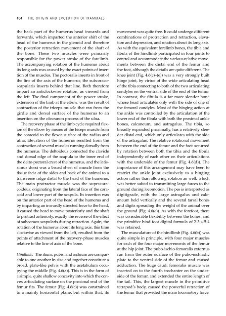
Hindlimb. <strong>The</strong> ilium, pubis, <strong>and</strong> ischium are comparable<br />
to one another in size <strong>and</strong> together constitute a<br />
broad, plate-like pelvis with the acetabulum occupying<br />
the middle (Fig. 4.6(a)). This is in the form <strong>of</strong><br />
a simple, quite shallow concavity into which the convex<br />
articulating surface on the proximal end <strong>of</strong> the<br />
femur fits. <strong>The</strong> femur (Fig. 4.6(c)) was constrained<br />
to a mainly horizontal plane, but within that, its<br />
movement was quite free. It could undergo different<br />
combinations <strong>of</strong> protraction <strong>and</strong> retraction, elevation<br />
<strong>and</strong> depression, <strong>and</strong> rotation about its long axis.<br />
As with the equivalent forelimb bones, the tibia <strong>and</strong><br />
fibula <strong>of</strong> the hindlimb participated in four joints to<br />
control <strong>and</strong> accommodate the various relative movements<br />
between the distal end <strong>of</strong> the femur <strong>and</strong><br />
the foot, although the details are quite different. <strong>The</strong><br />
knee joint (Fig. 4.6(c)–(e)) was a very strongly built<br />
hinge joint, by virtue <strong>of</strong> the wide articulating head<br />
<strong>of</strong> the tibia connecting to both <strong>of</strong> the two articulating<br />
condyles on the ventral side <strong>of</strong> the end <strong>of</strong> the femur.<br />
In contrast, the fibula is a far more slender bone<br />
whose head articulates only with the side <strong>of</strong> one <strong>of</strong><br />
the femoral condyles. Most <strong>of</strong> the hinging action at<br />
the ankle was controlled by the articulation <strong>of</strong> the<br />
lower end <strong>of</strong> the fibula with both the proximal ankle<br />
bones, calcaneum, <strong>and</strong> astragalus. <strong>The</strong> tibia, so<br />
broadly exp<strong>and</strong>ed proximally, has a relatively slender<br />
distal end, which only articulates with the side<br />
<strong>of</strong> the astragalus. <strong>The</strong> relative rotational movement<br />
between the end <strong>of</strong> the femur <strong>and</strong> the foot occurred<br />
by rotation between both the tibia <strong>and</strong> the fibula<br />
independently <strong>of</strong> each other on their articulations<br />
with the underside <strong>of</strong> the femur (Fig. 4.6(d)). <strong>The</strong><br />
importance <strong>of</strong> this arrangement may have been to<br />
restrict the ankle joint exclusively to a hinging<br />
action rather than allowing rotation as well, which<br />
was better suited to transmitting large forces to the<br />
ground during locomotion. <strong>The</strong> pes is interpreted as<br />
digitigrade, with the large astragalus <strong>and</strong> calcaneum<br />
held vertically <strong>and</strong> the several tarsal bones<br />
<strong>and</strong> digits spreading the weight <strong>of</strong> the animal over<br />
the ground (Fig. 4.6(e)). As with the forefoot, there<br />
was considerable flexibility between the bones, <strong>and</strong><br />
the primitive hind foot digital formula <strong>of</strong> 2-3-4-5-4<br />
was retained.<br />
<strong>The</strong> musculature <strong>of</strong> the hindlimb (Fig. 4.6(b)) was<br />
quite simple in principle, with four major muscles<br />
for each <strong>of</strong> the four major movements <strong>of</strong> the femur<br />
at the hip joint. <strong>The</strong> pubo-ischio-femoralis externus<br />
ran from the outer surface <strong>of</strong> the pubo-ischiadic<br />
plate to the ventral side <strong>of</strong> the femur <strong>and</strong> caused<br />
adduction. <strong>The</strong> huge caudi femoralis muscle was<br />
inserted on to the fourth trochanter on the underside<br />
<strong>of</strong> the femur, <strong>and</strong> extended the entire length <strong>of</strong><br />
the tail. This, the largest muscle in the primitive<br />
tetrapod’s body, caused the powerful retraction <strong>of</strong><br />
the femur that provided the main locomotory force.