

12th Congress of the European Hematology ... - Haematologica
12th Congress of the European Hematology ... - Haematologica
12th Congress of the European Hematology ... - Haematologica

You also want an ePaper? Increase the reach of your titles
YUMPU automatically turns print PDFs into web optimized ePapers that Google loves.
open-label, multi-centre study enrolled 35 patients (all provided informed<br />
consent) who were ei<strong>the</strong>r naïve or currently receiving anagrelide <strong>the</strong>rapy.<br />
For those on anagrelide prior to <strong>the</strong> study, <strong>the</strong> dosing regimen was<br />
maintained at <strong>the</strong> same pre-study level. For naïve patients, anagrelide<br />
was initiated at 0.5 mg daily. Patients were treated with anagrelide for<br />
3 months. Safety parameters and a range <strong>of</strong> PK and PD endpoints were<br />
assessed. Results. A total <strong>of</strong> 35 patients were enrolled in <strong>the</strong> study; 8 paediatric<br />
(″11 years) patients with essential thrombocythaemia (ET), 9 adolescents<br />
(age 12-15 years) with ET and 18 adolescents/adults (age ≥16<br />
years) with ET (12), polycy<strong>the</strong>mia vera (5) or ano<strong>the</strong>r MPD (1). The<br />
majority <strong>of</strong> <strong>the</strong> patients had experienced anagrelide prior to <strong>the</strong> study<br />
(94% in ≤15 years group, 72% in ≥16 years group) and exposure prior<br />
to <strong>the</strong> study was similar in <strong>the</strong> two groups. The median starting total daily<br />
dose was 1.0 mg in both groups, increasing to median final total daily<br />
doses <strong>of</strong> 1.5 mg and 2.0mg in <strong>the</strong> older and younger groups, respectively.<br />
The incidence <strong>of</strong> on-study AEs and related AEs in those aged ″15<br />
years (53% and 18%, respectively) was lower than for those in <strong>the</strong> ≥16<br />
years group (67% and 34%). There were no apparent differences in <strong>the</strong><br />
types <strong>of</strong> AEs observed in <strong>the</strong> two groups and no patients discontinued<br />
<strong>the</strong> study due to AEs. The three most frequent AEs were palpitations,<br />
fatigue and headache. The majority <strong>of</strong> events were considered drugrelated.<br />
There were no clinically significant abnormalities in terms <strong>of</strong><br />
24-hour ECGs, 12-lead ECGs or cardiovascular function (as assessed by<br />
echocardiogram). A comparison <strong>of</strong> PK data (normalised to 1mg dose and<br />
70kg bodyweight) showed that exposure to anagrelide (maximum<br />
observed plasma concentration [Cmax] and area under <strong>the</strong> plasma concentration-time<br />
curve during <strong>the</strong> dosing interval [AUCt]) was substantially<br />
lower (48% and 55%, respectively) in those ≤15 years than in those<br />
?16 years. No differences in <strong>the</strong> exposure to <strong>the</strong> active metabolite<br />
BCH24426 were observed. A number <strong>of</strong> correlations were analysed, and<br />
for example, when <strong>the</strong> age groups were analysed toge<strong>the</strong>r, <strong>the</strong>re was a<br />
good correlation between anagrelide and BCH24426 daily plasma exposure<br />
(Cmax and AUCt) and decreases in platelet count. Conclusions. The<br />
safety findings were as expected and were consistent with <strong>the</strong> pharmacological<br />
pr<strong>of</strong>ile <strong>of</strong> anagrelide and underlying diseases. Acceptable differences<br />
in PK pr<strong>of</strong>iles were observed and from this study current dosing<br />
regimens for anagrelide appear appropriate for use in children.<br />
0644<br />
PROGNOSTIC FACTORS IN IDIOPATHIC MYELOFIBROSIS: SINGLE CENTER ANALYSIS AND<br />
PROPOSAL FOR A NEW SCORING SYSTEM<br />
C.G. Gabutti, E.M. Pungolino, M.G. Draisci, M. Nichelatti, A. Nosari,<br />
S. Cantoni, G. Nador, M. Riva, E. Morra<br />
Ospedale Niguarda Ca'Granda, MILANO, Italy<br />
The survival <strong>of</strong> patients with IM is known to be extremely variable,<br />
ranging from a few months to more than 20 years. Such variability<br />
requires some prognostic factors, in order to orientate <strong>the</strong> <strong>the</strong>rapy choice<br />
in this cohort <strong>of</strong> patients. The Lille scoring system, proposed for patient<br />
risk stratification, seems unable to clearly discriminate between intermediate<br />
and high risk patients.<br />
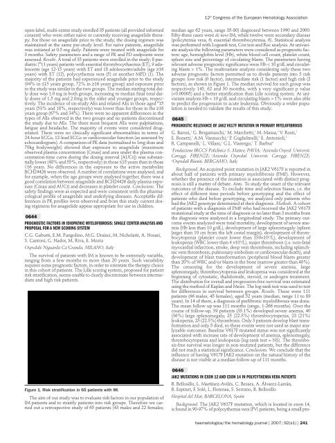
Figure 1. Risk stratification in 65 patients with IM.<br />
The aim <strong>of</strong> our study was to evaluate risk factors in our population <strong>of</strong><br />
IM patients and to stratify patients into risk groups. Therefore we carried<br />
out a retrospective study <strong>of</strong> 65 patients (43 males and 22 females;<br />
12 th <strong>Congress</strong> <strong>of</strong> <strong>the</strong> <strong>European</strong> <strong>Hematology</strong> Association<br />
median age 62 years, range 35-80) diagnosed between 1990 and 2000.<br />
Fifty-three cases were de novo IM, while twelve were secondary disease<br />
(policytemia vera, 6; essential thrombocytemia, 6). Statistical analysis<br />
was performed with Logrank test, Cox test and Roc analysis. At univariate<br />
analysis <strong>the</strong> following parameters were considered as prognostic factors:<br />
age, hemoglobin level (Hb), white blood cell count, platelet count,<br />
spleen size and percentage <strong>of</strong> circulating blasts. The parameters having<br />
relevant adverse prognostic significance were Hb < 10 g/dL and circulating<br />
blasts > 1%. The multivariate analysis considering only <strong>the</strong>se two<br />
adverse prognostic factors permitted us to divide patients into 3 risk<br />
groups: low risk (0 factor), intermediate risk (1 factor) and high risk (2<br />
factors), as shown in Figure 1. The median survival for each group was<br />
respectively 145, 62 and 50 months, with a very significant p value<br />
( 1% were also able<br />
to predict <strong>the</strong> progression to acute leukemia. Obviously a wider population<br />
is needed to validate <strong>the</strong> results <strong>of</strong> this study.<br />
0645<br />
PROGNOSTIC RELEVANCE OF JAK2 V617F MUTATION IN PRIMARY MYELOFIBROSIS<br />
G. Barosi, 1 G. Bergamaschi, 1 M. Marchetti, 1 M. Massa, 1 V. Rosti, 1<br />
E. Bonetti, 1 A.M. Vannucchi, 2 P. Guglielmelli, 2 E. Antonioli, 3<br />
R. Campanelli, 1 L. Villani, 1 G.L. Viarengo, 1 T. Barbui4 1 Fondazione IRCCS Policlinico S. Matteo, PAVIA; 2 Azienda Osped. Universit.<br />
Careggi, FIRENZE; 3 Azienda Ospedal. Universit. Careggi, FIRENZE;<br />
4 Ospedali Riuniti, BERGAMO, Italy<br />
Background. An acquired point mutation in JAK2 V617F is reported in<br />
about half <strong>of</strong> patients with primary myel<strong>of</strong>ibrosis (PMF). However,<br />
whe<strong>the</strong>r <strong>the</strong> presence <strong>of</strong> <strong>the</strong> mutation is associated with distinct prognosis<br />
is still a matter <strong>of</strong> debate. Aims. To study <strong>the</strong> onset <strong>of</strong> <strong>the</strong> relevant<br />
outcomes <strong>of</strong> <strong>the</strong> disease. To exclude time and selection biases, i.e. <strong>the</strong><br />
effect <strong>of</strong> different time periods before genotyping and <strong>the</strong> effect <strong>of</strong><br />
patients who died before genotyping, we analyzed only patients who<br />
had <strong>the</strong> JAK2 genotype determined at <strong>the</strong>ir diagnosis. Methods. A cohort<br />
<strong>of</strong> patients with a diagnosis <strong>of</strong> PMF who had received <strong>the</strong> JAK2 V617F<br />
mutational study at <strong>the</strong> time <strong>of</strong> diagnosis or no later than 3 months from<br />
<strong>the</strong> diagnosis were analyzed in a longitudinal study. The primary outcome<br />
events analyzed were total mortality, development <strong>of</strong> severe anemia<br />
(Hb less than 10 g/dL), development <strong>of</strong> large splenomegaly (spleen<br />
larger than 10 cm from <strong>the</strong> left costal margin), development <strong>of</strong> thrombocytopenia<br />
(platelet count lower than 150×10 9 /L), development <strong>of</strong><br />
leukopenia (WBC lower than 4 ×10 9 /L), major thrombosis (i.e. non-fatal<br />
myocardial infarction, stroke, deep vein thrombosis, including splanchnic<br />
vein thrombosis, pulmonary embolism or cardiovascular death), and<br />
development <strong>of</strong> blast transformation (peripheral blood blasts greater<br />
than 20% <strong>of</strong> WBC and/or blasts in <strong>the</strong> bone marrow greater than 40%).<br />
The censoring for <strong>the</strong> development <strong>of</strong> severe anemia, large<br />
splenomegaly, thrombocytopenia and leukopenia was considered at <strong>the</strong><br />
beginning <strong>of</strong> cytostatic, thalidomide, steroid, or androgen treatment.<br />
The distribution for overall and progression-free survival was estimated<br />
using <strong>the</strong> method <strong>of</strong> Kaplan and Meier. The log-rank test was used to test<br />
for differences in survival between groups. Results. These were 111<br />
patients (66 males, 45 females), aged 52 years (median, range 11 to 89<br />
years). In 14 <strong>of</strong> <strong>the</strong>m, a diagnosis <strong>of</strong> prefibrotic myel<strong>of</strong>ibrosis was done.<br />
The mean follow up was 111 months (range, 1-266 months). Over <strong>the</strong><br />
course <strong>of</strong> follow-up, 39 patients (35.1%) developed severe anemia, 40<br />
(36%) large splenomegaly, 25 (22.5%) thrombocytopenia, 23 (21%)<br />
leukopenia, 25 (22.5%) thrombosis. Only 3 patients develop blast transformation<br />
and only 5 died, so <strong>the</strong>se events were not used as major analyzable<br />
outcomes. Baseline V617F mutated status was not significantly<br />
associated with increase rate <strong>of</strong> development <strong>of</strong> anemia, splenomegaly,<br />
thrombocytopenia and leukopenia (log-rank test = NS). The thrombosis-free<br />
survival was longer in non-mutated patients, but <strong>the</strong> difference<br />
did not reach a statistical significance. Conclusions. We conclude that <strong>the</strong><br />
influence <strong>of</strong> having V617F JAK2 mutation on <strong>the</strong> natural history <strong>of</strong> <strong>the</strong><br />
disease is not visible at a median follow-up <strong>of</strong> 111 months.<br />
0646<br />
JAK2 MUTATIONS IN EXON 12 AND EXON 14 IN POLYCYTHEMIA VERA PATIENTS<br />
B. Bellosillo, L. Martínez-Avilés, C. Besses, A. Álvarez-Larrán,<br />
B. Espinet, F. Solé, L. Florensa, S. Serrano, B. Bellosillo<br />
Hospital del Mar, BARCELONA, Spain<br />
Background. The JAK2 V617F mutation, which is located in exon 14,<br />
is found in 90-97% <strong>of</strong> polycy<strong>the</strong>mia vera (PV) patients, being a small pro-<br />
haematologica/<strong>the</strong> hematology journal | 2007; 92(s1) | 241


