

12th Congress of the European Hematology ... - Haematologica
12th Congress of the European Hematology ... - Haematologica
12th Congress of the European Hematology ... - Haematologica

Create successful ePaper yourself
Turn your PDF publications into a flip-book with our unique Google optimized e-Paper software.
12 th <strong>Congress</strong> <strong>of</strong> <strong>the</strong> <strong>European</strong> <strong>Hematology</strong> Association<br />
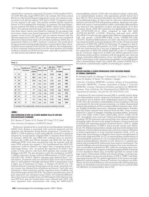
uated by flow-cytometry, expressed CD2 and/or CD25 (median 0.041%<br />
<strong>of</strong> CD45 + BM cells, range 0.004-0.28%). By contrast, MCs from normal<br />
BM (n=2), o<strong>the</strong>r hematological malignancies (n=4), and cutaneous mastocytosis<br />
(n=4) did not express CD2 and/or CD25. Cytogenetic analysis<br />
was normal in all cases. One patient revealed typical cutaneous<br />
lesions <strong>of</strong> CM (confirmed by histology). In summary, <strong>the</strong> final diagnosis<br />
was indolent SM (ISM) according to WHO criteria in 8/10 patients<br />
(80%). Five patients had both major and minor criteria <strong>of</strong> SM, whereas<br />
only three minor criteria were found in 3 patients. In one patient only<br />
two minor criteria were found (expression <strong>of</strong> CD25/CD2 on MC and<br />
tryptase serum levels >20 ng/mL), and one patient showed only <strong>the</strong><br />
aberrant expression CD25/CD2 on MC: <strong>the</strong>se patients were considered<br />
affected by Monoclonal Mast Cell activation syndrome (MMCAS), as<br />
proposed by Valent et al. (2007). Summary / Conclusions. Our results show<br />
strong association among anaphylaxis due to hymenoptera bites, abnormal<br />
basal serum tryptase levels and SM. In addition, <strong>the</strong> multiparametric<br />
flow cytometry analysis seems to be <strong>the</strong> most sensitive and reliable<br />
method to identify occult BM involvement, especially in patients without<br />
skin lesions and indolent disease.<br />
Table 1.<br />
1061<br />
HIGH EXPRESSION OF APAF-1XL IN BONE MARROW CELLS OF LOW RISK<br />
MYELODYSPLASTIC SYNDROME<br />
B.D. Benites, F. Traina, A.S.S. Duarte, F.F. Costa, S.T.O. Saad<br />
State University <strong>of</strong> Campinas, CAMPINAS, Brazil<br />
Background. Apoptosis has a crucial role in myelodysplastic syndromes<br />
(MDS). Early disease is associated with excessive apoptosis and <strong>the</strong><br />
apoptotic rate diminishes during disease progression. Cytochrome c/<br />
APAF-1/ CASP-9 pathway is <strong>the</strong> main pathway involved in apoptosis<br />
initiation by several stimuli. Original APAF-1 comprises three functional<br />
domains; APAF-1XL and APAF-1LN is<strong>of</strong>orms have an insertion<br />
between <strong>the</strong> CARD and ATPase domains and APAF-1XL also has an<br />
additional WDR. It has been reported that only <strong>the</strong> is<strong>of</strong>orms with <strong>the</strong><br />
extra WDR activate pro-caspase 9. We hypo<strong>the</strong>sized that APAF-1XL<br />
expression could be related to <strong>the</strong> higher rates <strong>of</strong> apoptosis found in<br />
early-stage disease. Aims. To analyse <strong>the</strong> expression <strong>of</strong> APAF-1 transcripts<br />
in bone marrow cells from MDS patients and correlate <strong>the</strong>se<br />
findings with IPSS. We also attempt to verify <strong>the</strong> modulation <strong>of</strong> APAF-<br />
1 mRNA expression during erythroid differentiation <strong>of</strong> CD34 + normal<br />
and MDS cells. Methods. Marrow aspirates were obtained from 7 normal<br />
donors, 14 patients with low risk MDS (IPSS: 9 low and 5 intermediate-1),<br />
and 8 patients with high risk disease (IPSS: 5 intermediate-2 and<br />
3 high), out <strong>of</strong> treatment (11 males, 11 females; 69 [40-91] yo). The<br />
National Ethical Committee Board approved <strong>the</strong> study; informed-written<br />
consent was obtained from all patients and donors. Total cells were<br />
submitted to RNA extraction and <strong>the</strong> expression level <strong>of</strong> mRNA was<br />
detected through real time RT-PCR. The relative quantification value <strong>of</strong><br />
gene expression was calculated using 2-DDct. For erythroid differentiation,<br />
bone marrow samples from one MDS patient with refractory anemia<br />
(IPSS low) and one normal donor were collected and CD34 + cells<br />
were separated from mononuclear cells using <strong>the</strong> MIDI-MACS<br />
392 | haematologica/<strong>the</strong> hematology journal | 2007; 92(s1)<br />
immunoaffinity columns. CD34 + cells were plated on plastic culture dishes<br />
in methylcellulose medium with appropriate growth factors for 6<br />
days. BFU-E, CFU-E and proerythroblasts were <strong>the</strong>n cultured in α MEM<br />
for an additional 8 days. At days 6 and 14, cells were collected and submitted<br />
to real time RT-PCR and apoptosis analysis. Apoptosis was quantified<br />
with anexin V and propidium iodide; erythroblast differentiation<br />
was observed with transferrin receptor and glycophorin A by flow<br />
cytometry. Results. APAF-1XL expression was significantly higher in low<br />
risk (15.327[3.095-121.1]) when compared to high risk MDS<br />
(4.379[2.412-44.632]; p=0.0103, Wilcoxon rank-sum test). APAF-<br />
1XL/APAF-1LN ratio was also significantly different comparing low and<br />
high risk groups (1.614[0.6598-6.964] versus 0.8904[0.155-2.99],<br />
p=0.0197). Erythroid differentiation <strong>of</strong> CD34 + cells from low risk MDS<br />
was characterized by increased apoptosis (20% at day 14) and increased<br />
expression <strong>of</strong> APAF-1XL mRNA (six fold at day 14 compared to day 6).<br />
In contrast, erythroid differentiation <strong>of</strong> CD34 + normal hematopoietic<br />
cells was characterized by a low rate <strong>of</strong> apoptosis (8% at day 14) and<br />
low expression <strong>of</strong> APAF-1XL mRNA (half fold on day 14 compared to<br />
day 6). Conclusions. High levels <strong>of</strong> APAF-1XL mRNA expression in low<br />
risk disease and its positive correlation with <strong>the</strong> apoptotic rate, observed<br />
during erythroblast differentiation <strong>of</strong> low risk MDS cells, suggest that<br />
APAF-1 participates in <strong>the</strong> augmented susceptibility <strong>of</strong> myelodysplastic<br />
cells to intramedullary death, since APAF-1XL, instead <strong>of</strong> APAF-1LN, is<br />
<strong>the</strong> is<strong>of</strong>orm constitutionally capable <strong>of</strong> activating pro-caspase 9.<br />
1062<br />
MYELOID SARCOMA: A CLINICO-PATHOLOGICAL STUDY INCLUDING IMAGING<br />
OF STROMAL COMPONENTS<br />
H. Schmitt-Graeff, 1 M. Metzger, 1 F. Feuerhake, 2 E.J. Juttner, 1 S. Haxelmans,<br />
3 B. Strahm, 4 H. Bertz, 5 M. Lübbert, 5 J. Burger6 1 University <strong>of</strong> Freiburg, FREIBURG, Germany; 2 Institute <strong>of</strong> Neuropathology,<br />
Universityh, FREIBURG, Germany; 3 Department <strong>of</strong> Biology I, University,<br />
FREIBURG, Germany; 4 Department <strong>of</strong> Pediatrics and Adolescent, FREIBURG,<br />
Germany; 5 Dept. <strong>of</strong> Medicine, Div. <strong>Hematology</strong>/Oncol, FREIBURG, Germany;<br />
6 Department <strong>of</strong> Leukemia, Unit 428 The Uni, HOUSTON, USA<br />
Background.The term myeloid sarcoma (MS) is currently used to designate<br />
extramedullary tumors composed <strong>of</strong> immature myeloid cells. Aims.<br />
Our purpose was to analyse clinico-pathological aspects <strong>of</strong> a large series<br />
<strong>of</strong> MS. Since <strong>the</strong> homing to extramedullary tissues resulting in MS may<br />
be promoted by <strong>the</strong> local microenvironment, we fur<strong>the</strong>r characterized<br />
stromal cell populations <strong>of</strong> MS occurring in different anatomic sites. Methods.<br />
Paraffin-embedded specimens containing representative formalinfixed<br />
MS biopsy material were retrieved from our files. Tissue sections<br />
stained for a panel <strong>of</strong> antigens including myeloperoxidase, lysozyme,<br />
CD3, CD4, CD20, CD34, CD41, CD61,CD68, CD117, CD99, CD 56,<br />
TdT , hemoglobin were reviewed. Clinical and laboratory informations<br />
including CXCR4 expression and survival data were obtained. Additional<br />
imaging studies focused on <strong>the</strong> spatial localization <strong>of</strong> microvessels and<br />
<strong>of</strong> stromal cells expressing stromal cell-derived factor-1 (SDF-1), cellular<br />
retinol-binding protein-1 (CRBP-1) and α-smooth muscle actin (SMA).<br />
Results. We obtained representative samples and clinical informations from<br />
61 patients. MS developed simultaneously with bone marrow infiltration<br />
as initial manifestation <strong>of</strong> acute myeloid leukaemia (AML, n=11), as<br />
relapse after chemo<strong>the</strong>rapy or allogeneic bone marrow transplantation <strong>of</strong><br />
a previously diagnosed AML (n=12), as extramedullary transformation <strong>of</strong><br />
a myelodysplastic syndrome (n=11) or as blast phase disease in typical or<br />
atypical myeloproliferative disorders (n=16). Moreover, 11 patients presented<br />
with an isolated MS without any bone marrow involvement. The<br />
myeloid infiltrates showed phenotypic features <strong>of</strong> an acute myelomonocytic<br />
or monoblastic leukaemia in 48% while <strong>the</strong> remaining cases corresponded<br />
to various AML categories including megakaryoblastic leukaemia<br />
or were classified as blastic type, not fur<strong>the</strong>r specified. In contrast to <strong>the</strong><br />
large variety <strong>of</strong> phenotypic pr<strong>of</strong>iles and clinical settings, a common feature<br />
<strong>of</strong> all samples was an increased density <strong>of</strong> SDF-1, CRBP-1 and SMAexpressing<br />
stromal cells.The spatial organization <strong>of</strong> <strong>the</strong> stroma was highlighted<br />
by confocal imaging. Summary/Conclusions. According to our observations,<br />
<strong>the</strong> functional interaction <strong>of</strong> local stromal cells with myeloid population<br />
via chemokines and chemokine-receptors may be crucial for blast<br />
cell infiltration <strong>of</strong> extramedullary sites in MS.


