

12th Congress of the European Hematology ... - Haematologica
12th Congress of the European Hematology ... - Haematologica
12th Congress of the European Hematology ... - Haematologica

Create successful ePaper yourself
Turn your PDF publications into a flip-book with our unique Google optimized e-Paper software.
12 th <strong>Congress</strong> <strong>of</strong> <strong>the</strong> <strong>European</strong> <strong>Hematology</strong> Association<br />
portion <strong>of</strong> cases negative for this mutation. Recently, it has been<br />
described that JAK2V617F negative PV patients have mutations in exon<br />
12 <strong>of</strong> <strong>the</strong> JAK2 gene. Aims. To determine <strong>the</strong> JAK2 mutational status<br />
analysing both exon 12 and exon 14 in a series <strong>of</strong> PV patients. Patients<br />
and Methods. In 86 patients (44M/42F) from a single institution diagnosed<br />
with PV, at a median age <strong>of</strong> 62 years (range 25-87), analysis <strong>of</strong><br />
JAK2 exons 12 and 14 was performed by direct sequencing using granulocyte<br />
RNA. At <strong>the</strong> time <strong>the</strong> JAK2 analysis was performed, 24/86 PV<br />
patients were receiving <strong>the</strong>rapy with platelet-lowering agents: hydroxyurea±ASA<br />
(n=23); anagrelide±ASA (n=1). Twenty-eight patients only<br />
received ASA and 34 patients did not receive any specific treatment.<br />
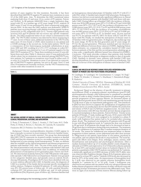
Results. The JAK2 mutational status <strong>of</strong> our cohort is summarized in <strong>the</strong><br />
Table. The V617F JAK2 mutation was detected by direct sequencing in<br />
77 cases. In two <strong>of</strong> <strong>the</strong>se cases, additional mutations to <strong>the</strong> V617F in <strong>the</strong><br />
same exon 14 were found: a C616C silent mutation in one patient and<br />
a C618R exchange in <strong>the</strong> second one. In this latter patient, V617F was<br />
a consequence <strong>of</strong> two heterozygous nucleotide substitutions at positions<br />
1849 and 1851 resulting in a GTC>TTT exchange at codon 617.<br />
Exon 12 mutations were detected in 3 out <strong>of</strong> 8 patients negative for <strong>the</strong><br />
JAK2 V617F mutation (37%). These three mutations in exon 12 were different<br />
from each o<strong>the</strong>r (Table 1). Moreover, exon 12 was analyzed in <strong>the</strong><br />
whole cohort, but no mutations were detected among JAK2V617F positive<br />
patients. In 5 patients no mutations were found in ei<strong>the</strong>r exon 12<br />
or exon 14. Conclusions. Mutations in exon 12 are detected in a percentage<br />
<strong>of</strong> JAK2V617F negative patients, but not in all cases. Exon 12 and<br />
exon 14 mutations are mutually exclusive, but <strong>the</strong> V617F mutation may<br />
coexist with o<strong>the</strong>r mutations in exon 14.<br />
Table 1.<br />
0647<br />
THE NATURAL HISTORY OF FAMILIAL CHRONIC MYELOPROLIFERATIVE DISORDERS:<br />
CLINICAL PRESENTATION, OUTCOME, AND ANTICIPATION<br />
E. Rumi, F. Passamonti, C. Elena, L. Arcaini, C. Del Curto, M.G. Della<br />
Porta, D. Pietra, E. Boveri, C. Pascutto, M. Cazzola, M. Lazzarino<br />
Fondazione IRCCS Policlinico San Matteo, PAVIA, Italy<br />
Background. Chronic myeloproliferative disorders (CMD) appear to<br />
have a sporadic occurrence in most instances. However, familial clustering<br />
<strong>of</strong> CMD has been recently reported. Our group has demonstrated<br />
that JAK2 (V617F) represents an acquired somatic mutation in familial<br />
CMD and that <strong>the</strong> mutation occurs as a secondary genetic event in <strong>the</strong><br />
background <strong>of</strong> a pre-existing clonal hematopoiesis. To date, no definite<br />
data are available on <strong>the</strong> clinical presentation and disease progression in<br />
patients with familial CMD. Aims. The aim <strong>of</strong> this study was to assess<br />
<strong>the</strong> clinical presentation and outcome <strong>of</strong> patients with familial CMD and<br />
to provide a comparison with sporadic cases. In addition, we studied <strong>the</strong><br />
phenomenon <strong>of</strong> anticipation in two-generation pairs. Patients and Methods.<br />
We interviewed on family history for CMD 348 patients with apparently<br />
sporadic CMD followed at our Department. Patients were grouped<br />
in two categories: familial cases, and sporadic cases as controls. Results.<br />
Among 348 patients, 30 pedigrees (8.6%) have been identified with one<br />
or more relatives affected with CMD. In familial cases, <strong>the</strong> diagnosis <strong>of</strong><br />
CMD in a member <strong>of</strong> <strong>the</strong> family did not prompt specific investigations<br />
within relatives. Pedigrees included 64 patients: 31 with polycy<strong>the</strong>mia<br />
vera (PV; 253 person-years <strong>of</strong> follow-up), 19 with essential thrombocy<strong>the</strong>mia<br />
(ET; 144 person-years <strong>of</strong> follow-up), and 14 with primary<br />
myel<strong>of</strong>ibrosis (PM; 84 person-years <strong>of</strong> follow-up). Nineteen families had<br />
242 | haematologica/<strong>the</strong> hematology journal | 2007; 92(s1)<br />
an homogeneous clinical phenotype (12 families with PV, 5 with ET, 2<br />
with PM), while 11 families had a mixed CMD phenotype. Kolmogorov-<br />
Smirnov test did not reveal statistically significant differences in clinical<br />
presentation between patients with familial CMD and those with sporadic<br />
CMD. During follow-up <strong>of</strong> familial CMD, <strong>the</strong> incidence <strong>of</strong> thrombosis<br />
was 26.8×1000 person-years (95% CI 12-60) in PV and 14.6×1000<br />
person-years (95% CI 3.7-58.7) in ET; <strong>the</strong> incidence <strong>of</strong> leukemia was 7.9<br />
×1000 person-years (95% CI 1.9-31.7) in PV and 48.6×1000 person-years<br />
(95% CI 18.2-129.5) in PM; <strong>the</strong> incidence <strong>of</strong> secondary myel<strong>of</strong>ibrosis<br />
was 4×1000 person-years (95% CI 0.5-28.4) in PV and 10.1×1000 person-years<br />
(95% CI 3.5-56.3) in ET. In familial cases, 10-year survival<br />
was 91.5% for patients with PV, 100% for those with ET and 30% for<br />
those with PM. Finally, we studied <strong>the</strong> anticipation <strong>of</strong> disease onset in<br />
15 families with two generation pairs. At diagnosis, <strong>the</strong> median age was<br />
61 years (range, 43-78) for <strong>the</strong> first generation and 37 years (range 23-<br />
57) for <strong>the</strong> second generation. Wilcoxon matched pair test showed a<br />
significant difference between <strong>the</strong>se values (p=0.0004). Applying Nelson<br />
Aalen estimator, we compared <strong>the</strong> cumulative hazard <strong>of</strong> CMD onset<br />
between patients <strong>of</strong> <strong>the</strong> first and <strong>the</strong> second generation adopting age as<br />
a time scale: a significantly different hazard was obtained (p=0.00001).<br />
Conclusions. This study provides evidence that patients with familial<br />
CMD have a clinical phenotype at diagnosis similar to that <strong>of</strong> sporadic<br />
CMD. Similarly to sporadic cases, patients with familial CMD may<br />
develop thrombosis or may progress to myel<strong>of</strong>ibrosis or leukemia. Our<br />
data are in favour <strong>of</strong> <strong>the</strong> anticipation <strong>of</strong> disease onset in familial CMD.<br />
0648<br />
CLINICAL AND MOLECULAR RESPONSE DURING PEGYLATED INTERFERON ALPHA<br />
THERAPY IN PRIMARY AND POST-POLYCYTHEMIC MYELOFIBROSIS<br />
H. Gisslinger, 1 B. Gisslinger, 1 M. Griesshammer, 2 C. Langer, 2 M. Fiegl, 3<br />
J. Thaler, 4 R. Schoder, 1 S. Schauer, 2 L. Muellauer, 1 I. Simonitsch-Klupp, 1<br />
R. Kralovics1 1 Medical University <strong>of</strong> Vienna, VIENNA; 2 Department <strong>of</strong> Medicine III, ULM,<br />
Germany; 3 Medical Unviversity <strong>of</strong> Innsbruck, INNSBRUCK, Austria;<br />
4 Klinikum Kreuzschwestern Wels, WELS, Austria<br />
Background. Based on <strong>the</strong> absence <strong>of</strong> specific treatment in primary<br />
myel<strong>of</strong>ibrosis (PMF) or postpolycy<strong>the</strong>mic myel<strong>of</strong>ibrosis (post-PV-MF)<br />
myelosuppressive <strong>the</strong>rapy with hydroxyurea is considered as standard<br />
<strong>the</strong>rapy in order to delay a fur<strong>the</strong>r deterioration <strong>of</strong> splenomegaly and to<br />
prevent progression <strong>of</strong> bone marrow fibrosis. An increased release <strong>of</strong><br />
platelet derived growth factor (PDGF) and transforming growth factor β<br />
(TGF-β) seem to play an importatant pathogenetic role. Aims.Interferon<br />
α (IFN) has been shown to suppress <strong>the</strong> proliferation <strong>of</strong> megakaryocyte<br />
cell lines and in vivo studies have shown that IFN inhibits <strong>the</strong> production<br />
and release <strong>of</strong> PDGF and TGF-β from megakaryocytes. An early <strong>the</strong>rapeutic<br />
intervention with IFN may <strong>the</strong>refore prevent disease progression<br />
to myeloid metaplasia in <strong>the</strong>se patients. Patients/Methods. Twenty-seven<br />
patients (10 female, 17 male; median age 58 years, 34-81 years) with PMF<br />
(n=25) or post-PV-MF (n=2) were assigned to be included in a phase II<br />
study with PegIntron.One patient had to be excluded after screening<br />
phase. Results. The treatment was initiated in 25 patients with 50 µg and<br />
in one with 80 µg PegIntron/week. In 7 patients <strong>the</strong> dose was increased<br />
to 80 µg/week. In all patients this dosage had to be decreased at least to<br />
50 µg/week due to intolerance during long-term treatment. The treatment<br />
had to be withdrawn in 6 patients after a median treatment duration <strong>of</strong><br />
24 weeks. The reason for withdrawal was treatment failure or disease<br />
progression in three patients, severe abdominal pain after dose increase<br />
to 100µg/week in one patient, heart failure in one patient, psychosis in<br />
one patient, and one patient did decide to stop treatment. The most frequent<br />
side effects were fatigue, fever, arthralgia, nausea, anemia, mild<br />
thrombopenia and leukopenia and psychological alterations. After a<br />
median treatment follow-up <strong>of</strong> 12 months platelet counts dropped significantly<br />
(p


