Timing, hosts and locations of (grouped) events of NanoImpactNet
Timing, hosts and locations of (grouped) events of NanoImpactNet
Timing, hosts and locations of (grouped) events of NanoImpactNet

Create successful ePaper yourself
Turn your PDF publications into a flip-book with our unique Google optimized e-Paper software.
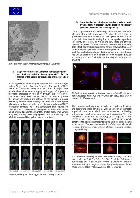
High Resolution Electron Microscopy image <strong>of</strong> ZnO particles<br />
2) Single Photon Emission Computed Tomography (SPECT)<br />
<strong>and</strong> Positron Emission Tomography (PET) for the<br />
analysis <strong>of</strong> the uptake, distribution <strong>and</strong> release <strong>of</strong> NPs in<br />
vivo.<br />
At the organism level, we propose the novel use <strong>of</strong> methodologies<br />
such as Single Photon Emission Computed Tomography (SPECT)<br />
<strong>and</strong> Positron Emission Tomography (PET). Both techniques allow<br />
for the three dimensional mapping or imaging <strong>of</strong> organs <strong>and</strong><br />
functional processes in the body through the detection <strong>of</strong><br />
radioactive species. SPECT <strong>and</strong> PET will be used to directly follow<br />
the uptake, distribution <strong>and</strong> release <strong>of</strong> the particles in animal<br />
models by different ingestion ways. To perform this task, special<br />
NPs have to be designed with tracers <strong>of</strong> gamma radiation (SPECT)<br />
or positron emitters (PET). This complicated task requires the<br />
fabrication <strong>and</strong> stabilization <strong>of</strong> these particles under conditions <strong>of</strong><br />
hot chemistry, taking into account the limited decay time. Wholebody<br />
analysis using direct imaging techniques <strong>of</strong> potentially toxic<br />
NP distribution <strong>and</strong> kinetics has been accomplished.<br />
Image sequence <strong>of</strong> PET scanning for g-Al218O3 NPs by IV route<br />
NanoSafetyCluster - Compendium 2012<br />
3) Quantification <strong>and</strong> distribution studies at cellular level,<br />
by Ion Beam Microscopy (IBM), Electron Microscopy<br />
(EM) <strong>and</strong> Confocal Laser Scanning (CLSM).<br />
There is a pr<strong>of</strong>ound lack <strong>of</strong> knowledge concerning the amount <strong>of</strong><br />
NPs present in a cell for an applied NP dose. In other words, a<br />
quantitative relation between dose <strong>and</strong> uptake <strong>of</strong> NPs at both<br />
organ <strong>and</strong> cellular level is missing. The particle uptake depends on<br />
the activity <strong>of</strong> the cells, as well as the size, shape <strong>and</strong> physicochemical<br />
properties <strong>of</strong> the nanomaterial. Therefore, the absence <strong>of</strong><br />
dose-effect relationships represents a serious drawback for proper<br />
risk evaluation <strong>of</strong> special intracellular developed effects. At cellular<br />
level, the localization <strong>and</strong> quantification <strong>of</strong> metal <strong>and</strong> metal oxide<br />
NPs will be performed by Ion Beam Microscopy (IBM), Electron<br />
Microscopy (EM) <strong>and</strong> Confocal Laser Scanning Microscopy (CLSM<br />
or LSCM).<br />
A) Confocal laser scanning microscopy image <strong>of</strong> HepG2 cells after<br />
being incubated with CeO2 NPs for 24hrs. (B) HepG2 cells without<br />
exposure to NPs as control.<br />
IBM is a unique <strong>and</strong> very powerful technique capable <strong>of</strong> localizing<br />
<strong>and</strong> quantifying these particles as well as performing elemental<br />
map distributions inside cells. It does not require particle labelling<br />
<strong>and</strong> relatively thick specimens can be investigated. The IBM<br />
technique is based on the targeting <strong>of</strong> a sample with high<br />
energetic ions (with approximately 2-3 MeV energy), which<br />
penetrate the targeted sample interacting with the electrons <strong>and</strong><br />
nuclei present. This leads to an excitation <strong>of</strong> electron shells, which<br />
rearrange themselves under emission <strong>of</strong> electromagnetic radiation<br />
(X-rays <strong>and</strong> light).<br />
PIXE elemental mapping <strong>of</strong> A549 cells treated during 72 h with<br />
various NPs: A- ZnO; B – CeO2; C – TiOx; D - FeOx . Left images<br />
demonstrate the P distribution (yellow is maximum, black is<br />
minimum) <strong>and</strong> right images – overlapping <strong>of</strong> two elements in the<br />
cells: S (green) <strong>and</strong> NPs metal (Zn, Ce, Ti, <strong>and</strong> Fe) (purple).<br />
Compendium <strong>of</strong> Projects in the European NanoSafety Cluster 37