annual report annual report annual report annual report 2005
annual report annual report annual report annual report 2005
annual report annual report annual report annual report 2005

Create successful ePaper yourself
Turn your PDF publications into a flip-book with our unique Google optimized e-Paper software.
NUCLEAR TECHNOLOGIES AND METHODS 129<br />
[4]. Günzel R., Betzl M., Alphonsa I., Ganguly B., John<br />
P.I., Mukherjee S.: Surf. Coat. Technol., 112, 307-309<br />
(1999).<br />
[5]. Sartowska B., Piekoszewski J., Waliś L., Szymczyk W.,<br />
Stanisławski J., Nowicki L., Ratajczak R., Kopcewicz<br />
M., Kalinowska J., Barcz A., Prokert F.: Vacuum, 78,<br />
181-186 (<strong>2005</strong>).<br />
[6]. Gavriljuk V.G., Berns H.: High nitrogen steels. Structure,<br />
properties, manufacture, applications. Springer-<br />
-Verlag, Berlin Heilderberg 1999, 376 p.<br />
[7]. Williamson D.L., Wang L., Wie R., Wilbur P.J.: Mater.<br />
Lett., 9, 9, 302-308 (1990).<br />
UV IRRADIATION OF TRACK MEMBRANES AS A METHOD<br />
FOR OBTAINING THE NECESSARY VALUE OF BRITTLENESS<br />
FOR GOOD FRACTURES OF SAMPLES FOR SEM OBSERVATIONS<br />
Bożena Sartowska, Oleg Orelovitch 1/ , Andrzej Nowicki<br />
1/<br />
Flerov Laboratory of Nuclear Reactions, Joint Institute for Nuclear Research, Dubna, Russia<br />
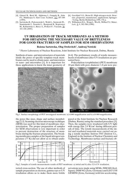
Synthesis of nano- and microstructures of materials<br />
inside the pores of specific template-track membranes<br />
can be used to obtain nano- and microwires<br />
or nano- and microtubes [1]. It is important for<br />
these applications to know the inner geometry of<br />
Fig.1. Surface morphology of PET investigated membrane: a) x1000 magnification and b) x10 000 magnification.<br />
Fig.2. Samples of tensile measurements results of PET membrane: a) initial and b) after 52 h UV irradiation.<br />
the pores like sizes, shape and surface morphology<br />
[2,3]. Scanning electron microscopy technique<br />
(SEM) was used for this kind of membrane characterisation.<br />
The proper preparation of samples<br />
for SEM observations is very important in order<br />
to prevent destruction of the structure of membrane<br />
during the fracture preparation. The breaking<br />
membranes samples at the liquid nitrogen temperature<br />
(77 K) did not allow us to obtain undistorted<br />
cross-section. The use of other methods of<br />
sample preparation as electron, gamma rays or UV<br />
irradiation allows us to make them more brittle<br />
[4-6]. The preliminary results of tensile measurements<br />
of membranes after UV irradiation are presented<br />
here.<br />
Poly(ethylene terephthalate) (PET) membrane<br />
10 µm thick with pore diameter 1.0 µm were prepared<br />
at the Joint Institute for Nuclear Research<br />
(Dubna, Russia) using the standard procedure [3].<br />
Then, the samples were irradiated with UV light<br />
with energy flux 2.8 W/cm 2 during different periods<br />
of time. The tensile measurements of the initial<br />
and irradiated materials were carried out using<br />
a tensile machine Instron 5565 (Instron Co.,<br />
England) in the Institute of Nuclear Chemistry and<br />
Technology. Membranes surface and fracture observations<br />
were made using SEMs: JSM 840 (Jeol,<br />
Japan), DSM 942 (Zeiss, Germany) and LEO 1530<br />
GEMINI (Zeiss, Germany) with low accelerating.