annual report annual report annual report annual report 2005
annual report annual report annual report annual report 2005
annual report annual report annual report annual report 2005

Create successful ePaper yourself
Turn your PDF publications into a flip-book with our unique Google optimized e-Paper software.
RADIOBIOLOGY 99<br />
DNA DAMAGE IN SUBFRACTIONS OF HUMAN LYMPHOCYTES<br />
IRRADIATED WITH LOW DOSES OF X-RADIATION<br />
Marcin Kruszewski 1,2/ , Maria Wojewódzka 1/ , Teresa Iwaneńko 1/ , Aneta Goździk 3/ ,<br />
Tomasz Ołdak 2,3/ , Eugeniusz K. Machaj 2,3/ , Zygmunt Pojda 2,3/<br />
1/<br />
Institute of Nuclear Chemistry and Technology, Warszawa, Poland<br />
2/<br />
The Maria Skłodowska-Curie Memorial Cancer Center and Institute of Oncology, Warszawa, Poland<br />
3/<br />
Military Institute of Hygiene and Epidemiology, Warszawa, Poland<br />
The aim of this study was to find a reliable, sensitive,<br />
fast and relatively cheap method to estimate<br />
radiation-induced DNA damage in human lymphocytes<br />
during a short time period after irradiation.<br />
We compared three methods: comet assay,<br />
micronucleus test and formation of phosphorylated<br />
histone H2AX foci.<br />
Human peripheral blood lymphocytes were<br />
collected by vein puncture and fractionated using<br />
T Cell Isolation Kit II and B Cell Isolation Kit II<br />
(Miltenyi Biotec), according to the manufacturer<br />
recommendations. Homogeneity of both T and B<br />
cell populations was 97-99%, as estimated by flow<br />
cytometry. Cells were irradiated with low doses of<br />
X-rays (0-1 Gy, 180 kV, 18 mA, 1 mm Cu filter)<br />
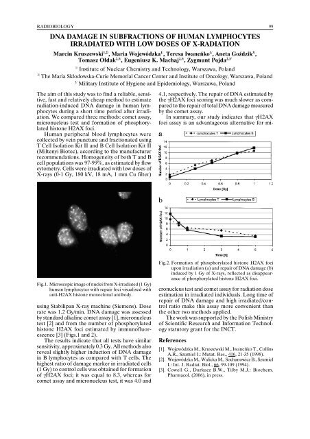
Fig.1. Microscopic image of nuclei from X-irradiated (1 Gy)<br />
human lymphocytes with repair foci visualised with<br />
anti-H2AX histone monoclonal antibody.<br />
using Stabilipan X-ray machine (Siemens). Dose<br />
rate was 1.2 Gy/min. DNA damage was assessed<br />
by standard alkaline comet assay [1], micronucleus<br />
test [2] and from the number of phosphorylated<br />
histone H2AX foci estimated by immunofluorescence<br />
[3] (Figs.1 and 2).<br />
The results indicate that all tests have similar<br />
sensitivity, approximately 0.3 Gy. All methods also<br />
reveal slightly higher induction of DNA damage<br />
in B lymphocytes as compared with T cells. The<br />
highest ratio of damage marker in irradiated cells<br />
(1 Gy) to control cells was obtained for formation<br />
of γH2AX foci; it was equal to 8.3, whereas for<br />
comet assay and micronucleus test, it was 4.0 and<br />
Fig.2. Formation of phosphorylated histone H2AX foci<br />
upon irradiation (a) and repair of DNA damage (b)<br />
induced by 1 Gy of X-rays, reflected as disappearance<br />
of phosphorylated histone H2AX foci.<br />
4.1, respectively. The repair of DNA estimated by<br />
the γH2AX foci scoring was much slower as compared<br />
to the repair of total DNA damage measured<br />
by the comet assay.<br />
In summary, our study indicates that γH2AX<br />
foci assay is an advantageous alternative for micronucleus<br />
test and comet assay for radiation dose<br />
estimation in irradiated individuals. Long time of<br />
repair of DNA damage and high irradiated/control<br />
ratio make this assay more convenient than<br />
the other two methods applied.<br />
The work was supported by the Polish Ministry<br />
of Scientific Research and Information Technology<br />
statutory grant for the INCT.<br />
References<br />
[1]. Wojewódzka M., Kruszewski M., Iwaneńko T., Collins<br />
A.R., Szumiel I.: Mutat. Res., 416, 21-35 (1998).<br />
[2]. Wojewódzka M., Walicka M., Sochanowicz B., Szumiel<br />
I.: Int. J. Radiat. Biol., 66, 99-109 (1994).<br />
[3]. Cowell G., Durkacz B.W., Tilby M.J.: Biochem.<br />
Pharmacol. (2006), in press.