

Segmentation of 3D Tubular Tree Structures in Medical Images ...
Segmentation of 3D Tubular Tree Structures in Medical Images ...
Segmentation of 3D Tubular Tree Structures in Medical Images ...

Create successful ePaper yourself
Turn your PDF publications into a flip-book with our unique Google optimized e-Paper software.
5.3. Evaluation and Results 81<br />
5.3.1 Phantom Vessel <strong>Tree</strong><br />
To evaluate the performance <strong>of</strong> our approach under the <strong>in</strong>fluence <strong>of</strong> vary<strong>in</strong>g noise levels,<br />
scan resolutions, contrast situations, and tube diameters, we produced a plastic phantom<br />
“vessel tree” with known digital ground truth. The design <strong>of</strong> the “vessel tree” (Fig. 5.2(a)),<br />
that was manufactured with a rapid prototyp<strong>in</strong>g mach<strong>in</strong>e, is <strong>in</strong>spired by the branch<strong>in</strong>g<br />
pattern <strong>of</strong> the human portal ve<strong>in</strong> tree <strong>of</strong> the liver and consists <strong>of</strong> about 600 cyl<strong>in</strong>drical<br />
branches with vary<strong>in</strong>g orientation and diameter (1 mm to 16 mm).<br />
The phantom was<br />
scanned with a Siemens Somatom Sensation 64 CT scanner with various resolutions to<br />
produce different noise levels, partial volume effects, and image reconstruction artifacts.<br />
To generate different contrast situations, different “backgrounds” were used: water and<br />
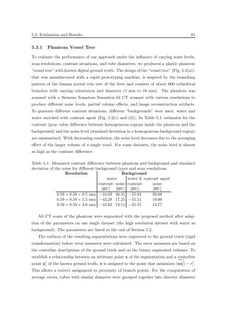
water enriched with contrast agent (Fig. 5.2(c) and (d)). In Table 5.1, estimates for the<br />
contrast (gray value difference between homogeneous regions <strong>in</strong>side the phantom and the<br />
background) and the noise level (standard deviation <strong>in</strong> a homogeneous background region)<br />
are summarized. With decreas<strong>in</strong>g resolution, the noise level decreases due to the averag<strong>in</strong>g<br />
effect <strong>of</strong> the larger volume <strong>of</strong> a s<strong>in</strong>gle voxel. For some datasets, the noise level is almost<br />
as high as the contrast difference.<br />
Table 5.1: Measured contrast difference between phantom and background and standard<br />
deviation <strong>of</strong> the noise for different background types and scan resolutions.<br />
Resolution<br />
Background<br />
water water & contrast agent<br />
contrast noise contrast noise<br />
[HU] [HU] [HU] [HU]<br />
0.59 × 0.59 × 0.5 mm −44.03 40.31 −55.83 39.68<br />
0.59 × 0.59 × 1.5 mm −43.28 17.25 −55.51 19.60<br />
0.59 × 0.59 × 3.0 mm −42.62 13.11 −55.57 14.77<br />
All CT scans <strong>of</strong> the phantom were segmented with the proposed method after adaption<br />
<strong>of</strong> the parameters on one s<strong>in</strong>gle dataset (the high resolution dataset with water as<br />
background). The parameters are listed at the end <strong>of</strong> Section 5.2.<br />
The surfaces <strong>of</strong> the result<strong>in</strong>g segmentations were registered to the ground truth (rigid<br />
transformation) before error measures were calculated. The error measures are based on<br />
the centerl<strong>in</strong>e descriptions <strong>of</strong> the ground truth and on the b<strong>in</strong>ary segmented volumes. To<br />
establish a relationship between an arbitrary po<strong>in</strong>t x <strong>of</strong> the segmentation and a centerl<strong>in</strong>e<br />
−→<br />
po<strong>in</strong>t x j i<br />
<strong>of</strong> the known ground truth, x is assigned to the po<strong>in</strong>t that m<strong>in</strong>imizes | xx<br />
j<br />
i | − rj i .<br />
This allows a correct assignment <strong>in</strong> proximity <strong>of</strong> branch po<strong>in</strong>ts. For the computation <strong>of</strong><br />
average errors, tubes with similar diameter were grouped together <strong>in</strong>to discrete diameter


