

Segmentation of 3D Tubular Tree Structures in Medical Images ...
Segmentation of 3D Tubular Tree Structures in Medical Images ...
Segmentation of 3D Tubular Tree Structures in Medical Images ...

You also want an ePaper? Increase the reach of your titles
YUMPU automatically turns print PDFs into web optimized ePapers that Google loves.
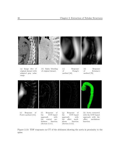
46 Chapter 2. Extraction <strong>of</strong> <strong>Tubular</strong> <strong>Structures</strong><br />
(a) Image slice <strong>of</strong><br />
orig<strong>in</strong>al dataset with<br />
adapted gray value<br />
range.<br />
(b) Alpha blend<strong>in</strong>g<br />
<strong>of</strong> orig<strong>in</strong>al dataset.<br />
(c) Response<br />
<strong>of</strong> Frangi’s<br />
method [44].<br />
(d) Response<br />
<strong>of</strong> Krissian’s<br />
method [70].<br />
(e) Response <strong>of</strong><br />
Pock’s method [114].<br />
(f) Response <strong>of</strong><br />
the GVF-based<br />
approach with<br />
the central medialness<br />
function<br />
(Section 2.3.1).<br />
(g) Response <strong>of</strong><br />
the GVF-based<br />
approach with<br />
the <strong>of</strong>fset medialness<br />
function<br />
(Section 2.3.2).<br />
(h) Aorta extracted<br />
with the GVF-based<br />
approach with the<br />
<strong>of</strong>fset medialness<br />
function.<br />
Figure 2.13: TDF responses on CT <strong>of</strong> the abdomen show<strong>in</strong>g the aorta <strong>in</strong> proximity to the<br />
sp<strong>in</strong>e.


