

Segmentation of 3D Tubular Tree Structures in Medical Images ...
Segmentation of 3D Tubular Tree Structures in Medical Images ...
Segmentation of 3D Tubular Tree Structures in Medical Images ...

Create successful ePaper yourself
Turn your PDF publications into a flip-book with our unique Google optimized e-Paper software.
52 Chapter 3. Group<strong>in</strong>g and L<strong>in</strong>kage <strong>in</strong>to Connected Networks<br />
obta<strong>in</strong>ed from this image gradient <strong>in</strong>formation. We used the GVF field already for identification<br />
<strong>of</strong> tubular objects as a replacement for the multi-scale gradient vector computation<br />
for detection <strong>of</strong> tubular structures (Section 2.3). In comb<strong>in</strong>ation with a tube likel<strong>in</strong>ess<br />
measure based on central or <strong>of</strong>fset-medialness functions, the overall approach allows detection<br />
<strong>of</strong> tubular objects at their centerl<strong>in</strong>es show<strong>in</strong>g several advantages compared to<br />
conventional multi-scale TDFs. But similar to other TDFs, the method does not produce<br />
valid centerl<strong>in</strong>e paths <strong>in</strong> the proximity <strong>of</strong> junctions or diseases like stenosis where<br />
the objects shapes strongly deviates from the typical tubular shape. However, the GVF<br />
shows another property that can be used to solve this problem and that has been used<br />
for extraction <strong>of</strong> curve skeletons (CS) from b<strong>in</strong>ary segmentations by the skeletonization<br />
approach <strong>of</strong> Hassouna and Farag [53, 54]. In contrast to skeletonization approaches that<br />
are based on the distance transformation, the magnitude <strong>of</strong> the GVF always decreases<br />
towards the medial curves <strong>in</strong>dependent <strong>of</strong> the object’s shape. This property <strong>of</strong> the GVF<br />
enables the extraction <strong>of</strong> the centerl<strong>in</strong>es also <strong>in</strong> case <strong>of</strong> junctions or plate like structures.<br />
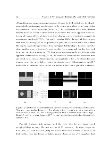
(a) (b) (c) (d) (e)<br />
Figure 3.2: Illustration <strong>of</strong> the basic idea on 2D cross section pr<strong>of</strong>iles <strong>of</strong> some <strong>3D</strong> structures.<br />
Top row: cross section T-junction <strong>of</strong> a tubular object; bottom row: structures with a<br />
circular cross section (left) and a strong deviation from a circular cross section (right).<br />
From left to right: orig<strong>in</strong>al dataset, GVF, derived tube-likel<strong>in</strong>ess, derived medialness λ(x),<br />
extracted CS.<br />
Fig. 3.2 illustrates this property and the basic idea for our image based<br />
group<strong>in</strong>g/l<strong>in</strong>kage on some 2D cross sections <strong>of</strong> <strong>3D</strong> structures. In these images the<br />
GVF field, the TDF response (us<strong>in</strong>g the central medialness function as described <strong>in</strong><br />
Section 2.3.1), and the derived medialness measure based on the GVF magnitude that


