

Segmentation of 3D Tubular Tree Structures in Medical Images ...
Segmentation of 3D Tubular Tree Structures in Medical Images ...
Segmentation of 3D Tubular Tree Structures in Medical Images ...

Create successful ePaper yourself
Turn your PDF publications into a flip-book with our unique Google optimized e-Paper software.
7.2. Methods 113<br />
Additionally, the extracted centerl<strong>in</strong>es are dilated and added to the segmentation result<br />
to assure 6-connected segmentation results.<br />
Parameters: The follow<strong>in</strong>g set <strong>of</strong> parameters is used for segmentation <strong>of</strong> the datasets:<br />
σ = 0.5mm, F max = 200, µ = 5 for the GVF us<strong>in</strong>g 500 iterations, t high = 0.5, t low = 0.1,<br />
t s = 5, and t m = 0.5.<br />
7.2.2 Airway <strong>Tree</strong> Reconstruction Based on Multi-Scale Tube Detection<br />
In this section, we present an automated approach for the reconstruction <strong>of</strong> airway trees.<br />
It is based no our general approach for segmentation <strong>of</strong> branched tubular networks as<br />
outl<strong>in</strong>ed <strong>in</strong> Section 1.2 to achieve a high robust aga<strong>in</strong>st local disturbances which can<br />
result from disease or imag<strong>in</strong>g artifacts, for example. The method consists <strong>of</strong> three ma<strong>in</strong><br />
process<strong>in</strong>g steps: preprocess<strong>in</strong>g, extraction <strong>of</strong> tubular objects, and a group<strong>in</strong>g and l<strong>in</strong>kage<br />
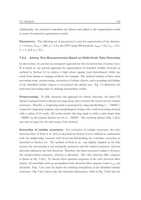
<strong>of</strong> the identified tubular objects to reconstruct the airway tree. Fig. 7.4 illustrates the<br />
<strong>in</strong>dividual process<strong>in</strong>g steps by show<strong>in</strong>g <strong>in</strong>termediate results.<br />
Preprocess<strong>in</strong>g: To fully automate the approach for airway detection, the <strong>in</strong>put CT<br />
dataset is preprocessed to discard non lung tissue and to restrict the search area for tubular<br />
structures. Therefor, a rough lung mask is generated by us<strong>in</strong>g threshold<strong>in</strong>g (< −700HU),<br />
connected component analysis, and morphological clos<strong>in</strong>g with a ball structur<strong>in</strong>g element<br />
with a radius <strong>of</strong> 10 voxels. All voxels outside this lung mask or with a value larger than<br />
−700HU <strong>in</strong> the orig<strong>in</strong>al dataset are set to −700HU. The result<strong>in</strong>g dataset (Fig. 7.4(a))<br />
was used as <strong>in</strong>put for the later steps <strong>of</strong> the method.<br />
Extraction <strong>of</strong> tubular structures: For extraction <strong>of</strong> tubular structures, the tube<br />
detection filter <strong>of</strong> Pock et al. [113] as described <strong>in</strong> Section 2.2.2 is utilized <strong>in</strong> comb<strong>in</strong>ation<br />
with the height-ridge traversal with hysteresis-threshold<strong>in</strong>g for centerl<strong>in</strong>e extraction as<br />
described <strong>in</strong> Section 2.4. The method <strong>of</strong> Pock et al. was slightly adapted as for th<strong>in</strong><br />
airways the surround<strong>in</strong>g is not necessarily symmetric and the utilized symmetry criterion<br />
adversely <strong>in</strong>fluences the tube detection. Therefore, for these structures (radius ≤ 0.5mm),<br />
the variance-based symmetry criterion is discarded. The tube detection filter response<br />
is shown <strong>in</strong> Fig. 7.4(b). To discard short spurious responses <strong>of</strong> the tube detection filter<br />
(noise), all centerl<strong>in</strong>es with an accumulated tube detection filter response below t conf are<br />
discarded. Figs. 7.4(c) and (d) depict the result<strong>in</strong>g descriptions <strong>of</strong> the identified tubular<br />
structures. Fig. 7.4(c) shows only the centerl<strong>in</strong>e <strong>in</strong>formation, while <strong>in</strong> Fig. 7.4(d) also the


