

Segmentation of 3D Tubular Tree Structures in Medical Images ...
Segmentation of 3D Tubular Tree Structures in Medical Images ...
Segmentation of 3D Tubular Tree Structures in Medical Images ...

You also want an ePaper? Increase the reach of your titles
YUMPU automatically turns print PDFs into web optimized ePapers that Google loves.
3.4. Experiments 61<br />
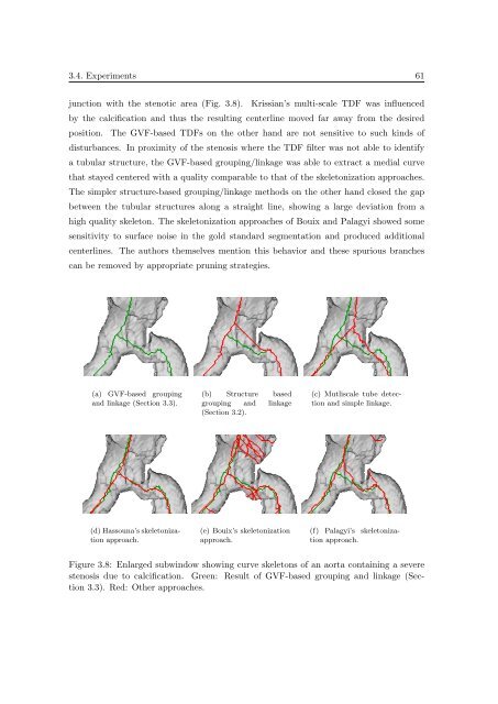
junction with the stenotic area (Fig. 3.8). Krissian’s multi-scale TDF was <strong>in</strong>fluenced<br />
by the calcification and thus the result<strong>in</strong>g centerl<strong>in</strong>e moved far away from the desired<br />
position. The GVF-based TDFs on the other hand are not sensitive to such k<strong>in</strong>ds <strong>of</strong><br />
disturbances. In proximity <strong>of</strong> the stenosis where the TDF filter was not able to identify<br />
a tubular structure, the GVF-based group<strong>in</strong>g/l<strong>in</strong>kage was able to extract a medial curve<br />
that stayed centered with a quality comparable to that <strong>of</strong> the skeletonization approaches.<br />
The simpler structure-based group<strong>in</strong>g/l<strong>in</strong>kage methods on the other hand closed the gap<br />
between the tubular structures along a straight l<strong>in</strong>e, show<strong>in</strong>g a large deviation from a<br />
high quality skeleton. The skeletonization approaches <strong>of</strong> Bouix and Palagyi showed some<br />
sensitivity to surface noise <strong>in</strong> the gold standard segmentation and produced additional<br />
centerl<strong>in</strong>es. The authors themselves mention this behavior and these spurious branches<br />
can be removed by appropriate prun<strong>in</strong>g strategies.<br />
(a) GVF-based group<strong>in</strong>g<br />
and l<strong>in</strong>kage (Section 3.3).<br />
(b) Structure based<br />
group<strong>in</strong>g and l<strong>in</strong>kage<br />
(Section 3.2).<br />
(c) Mutliscale tube detection<br />
and simple l<strong>in</strong>kage.<br />
(d) Hassouna’s skeletonization<br />
approach.<br />
(e) Bouix’s skeletonization<br />
approach.<br />
(f) Palagyi’s skeletonization<br />
approach.<br />
Figure 3.8: Enlarged subw<strong>in</strong>dow show<strong>in</strong>g curve skeletons <strong>of</strong> an aorta conta<strong>in</strong><strong>in</strong>g a severe<br />
stenosis due to calcification. Green: Result <strong>of</strong> GVF-based group<strong>in</strong>g and l<strong>in</strong>kage (Section<br />
3.3). Red: Other approaches.


