

Segmentation of 3D Tubular Tree Structures in Medical Images ...
Segmentation of 3D Tubular Tree Structures in Medical Images ...
Segmentation of 3D Tubular Tree Structures in Medical Images ...

Create successful ePaper yourself
Turn your PDF publications into a flip-book with our unique Google optimized e-Paper software.
3.4. Experiments 55<br />
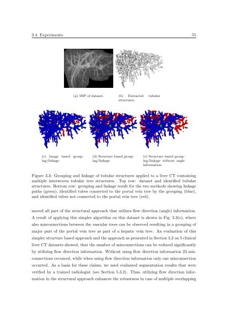
(a) MIP <strong>of</strong> dataset. (b) Extracted tubular<br />
structures.<br />
(c) Image based group<strong>in</strong>g/l<strong>in</strong>kage.<br />
(d) Structure based group<strong>in</strong>g/l<strong>in</strong>kage.<br />
(e) Structure based group<strong>in</strong>g/l<strong>in</strong>kage<br />
without angle<br />
<strong>in</strong>formation.<br />
Figure 3.3: Group<strong>in</strong>g and l<strong>in</strong>kage <strong>of</strong> tubular structures applied to a liver CT conta<strong>in</strong><strong>in</strong>g<br />
multiple <strong>in</strong>terwoven tubular tree structures. Top row: dataset and identified tubular<br />
structures. Bottom row: group<strong>in</strong>g and l<strong>in</strong>kage result for the two methods show<strong>in</strong>g l<strong>in</strong>kage<br />
paths (green), identified tubes connected to the portal ve<strong>in</strong> tree by the group<strong>in</strong>g (blue),<br />
and identified tubes not connected to the portal ve<strong>in</strong> tree (red).<br />
moved all part <strong>of</strong> the structural approach that utilizes flow direction (angle) <strong>in</strong>formation.<br />
A result <strong>of</strong> apply<strong>in</strong>g this simpler algorithm on this dataset is shown <strong>in</strong> Fig. 3.3(e), where<br />
also misconnections between the vascular trees can be observed result<strong>in</strong>g <strong>in</strong> a group<strong>in</strong>g <strong>of</strong><br />
major part <strong>of</strong> the portal ve<strong>in</strong> tree as part <strong>of</strong> a hepatic ve<strong>in</strong> tree. An evaluation <strong>of</strong> this<br />
simpler structure based approach and the approach as presented <strong>in</strong> Section 3.2 on 5 cl<strong>in</strong>ical<br />
liver CT datasets showed, that the number <strong>of</strong> misconnections can be reduced significantly<br />
by utiliz<strong>in</strong>g flow direction <strong>in</strong>formation. Without us<strong>in</strong>g flow direction <strong>in</strong>formation 23 misconnections<br />
occurred, while when us<strong>in</strong>g flow direction <strong>in</strong>formation only one misconnection<br />
occurred. As a basis for these claims, we used evaluated segmentation results that were<br />
verified by a tra<strong>in</strong>ed radiologist (see Section 5.3.2). Thus, utiliz<strong>in</strong>g flow direction <strong>in</strong>formation<br />
<strong>in</strong> the structural approach enhances the robustness <strong>in</strong> case <strong>of</strong> multiple overlapp<strong>in</strong>g


