

Segmentation of 3D Tubular Tree Structures in Medical Images ...
Segmentation of 3D Tubular Tree Structures in Medical Images ...
Segmentation of 3D Tubular Tree Structures in Medical Images ...

You also want an ePaper? Increase the reach of your titles
YUMPU automatically turns print PDFs into web optimized ePapers that Google loves.
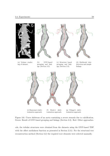
3.4. Experiments 59<br />
(a) Volume render<strong>in</strong>g<br />
<strong>of</strong> dataset.<br />
(b) GVF-based<br />
group<strong>in</strong>g and l<strong>in</strong>kage<br />
(Section 3.3).<br />
(c) Structure based<br />
group<strong>in</strong>g and l<strong>in</strong>kage<br />
(Section 3.2).<br />
(d) Mutliscale tube<br />
detection and simple<br />
l<strong>in</strong>kage.<br />
(e) Hassouna’s skeletonization<br />
approach.<br />
(f) Bouix’s skeletonization<br />
approach.<br />
(g) Palagyi’s skeletonization<br />
approach.<br />
Figure 3.6: Curve skeletons <strong>of</strong> an aorta conta<strong>in</strong><strong>in</strong>g a severe stenosis due to calcification.<br />
Green: Result <strong>of</strong> GVF-based group<strong>in</strong>g and l<strong>in</strong>kage (Section 3.3). Red: Other approaches.<br />
ods, the tubular structures were obta<strong>in</strong>ed from the datasets us<strong>in</strong>g the GVF-based TDF<br />
with the <strong>of</strong>fset medialness function as presented <strong>in</strong> Section 2.3.2. For the structural tree<br />
reconstruction method (Section 3.2) the required root elements were selected manually.


