

Segmentation of 3D Tubular Tree Structures in Medical Images ...
Segmentation of 3D Tubular Tree Structures in Medical Images ...
Segmentation of 3D Tubular Tree Structures in Medical Images ...

Create successful ePaper yourself
Turn your PDF publications into a flip-book with our unique Google optimized e-Paper software.
7.2. Methods 111<br />
shown <strong>in</strong> Fig. 7.1.<br />
(a) (b) (c) (d)<br />
(e) (f) (g) (h)<br />
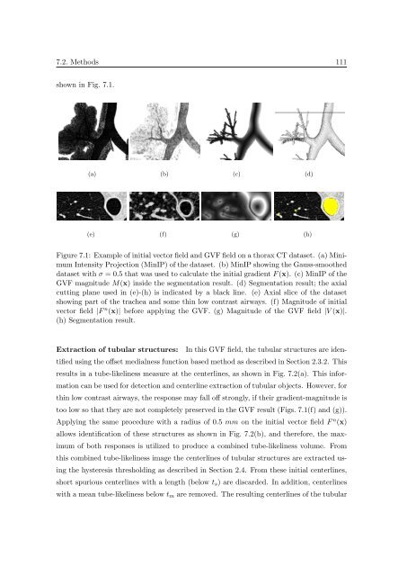
Figure 7.1: Example <strong>of</strong> <strong>in</strong>itial vector field and GVF field on a thorax CT dataset. (a) M<strong>in</strong>imum<br />
Intensity Projection (M<strong>in</strong>IP) <strong>of</strong> the dataset. (b) M<strong>in</strong>IP show<strong>in</strong>g the Gauss-smoothed<br />
dataset with σ = 0.5 that was used to calculate the <strong>in</strong>itial gradient F (x). (c) M<strong>in</strong>IP <strong>of</strong> the<br />
GVF magnitude M(x) <strong>in</strong>side the segmentation result. (d) <strong>Segmentation</strong> result; the axial<br />
cutt<strong>in</strong>g plane used <strong>in</strong> (e)-(h) is <strong>in</strong>dicated by a black l<strong>in</strong>e. (e) Axial slice <strong>of</strong> the dataset<br />
show<strong>in</strong>g part <strong>of</strong> the trachea and some th<strong>in</strong> low contrast airways. (f) Magnitude <strong>of</strong> <strong>in</strong>itial<br />
vector field |F n (x)| before apply<strong>in</strong>g the GVF. (g) Magnitude <strong>of</strong> the GVF field |V (x)|.<br />
(h) <strong>Segmentation</strong> result.<br />
Extraction <strong>of</strong> tubular structures: In this GVF field, the tubular structures are identified<br />
us<strong>in</strong>g the <strong>of</strong>fset medialness function based method as described <strong>in</strong> Section 2.3.2. This<br />
results <strong>in</strong> a tube-likel<strong>in</strong>ess measure at the centerl<strong>in</strong>es, as shown <strong>in</strong> Fig. 7.2(a). This <strong>in</strong>formation<br />
can be used for detection and centerl<strong>in</strong>e extraction <strong>of</strong> tubular objects. However, for<br />
th<strong>in</strong> low contrast airways, the response may fall <strong>of</strong>f strongly, if their gradient-magnitude is<br />
too low so that they are not completely preserved <strong>in</strong> the GVF result (Figs. 7.1(f) and (g)).<br />
Apply<strong>in</strong>g the same procedure with a radius <strong>of</strong> 0.5 mm on the <strong>in</strong>itial vector field F n (x)<br />
allows identification <strong>of</strong> these structures as shown <strong>in</strong> Fig. 7.2(b), and therefore, the maximum<br />
<strong>of</strong> both responses is utilized to produce a comb<strong>in</strong>ed tube-likel<strong>in</strong>ess volume. From<br />
this comb<strong>in</strong>ed tube-likel<strong>in</strong>ess image the centerl<strong>in</strong>es <strong>of</strong> tubular structures are extracted us<strong>in</strong>g<br />
the hysteresis threshold<strong>in</strong>g as described <strong>in</strong> Section 2.4. From these <strong>in</strong>itial centerl<strong>in</strong>es,<br />
short spurious centerl<strong>in</strong>es with a length (below t s ) are discarded. In addition, centerl<strong>in</strong>es<br />
with a mean tube-likel<strong>in</strong>ess below t m are removed. The result<strong>in</strong>g centerl<strong>in</strong>es <strong>of</strong> the tubular


