

Segmentation of 3D Tubular Tree Structures in Medical Images ...
Segmentation of 3D Tubular Tree Structures in Medical Images ...
Segmentation of 3D Tubular Tree Structures in Medical Images ...

Create successful ePaper yourself
Turn your PDF publications into a flip-book with our unique Google optimized e-Paper software.
114 Chapter 7. Airway <strong>Tree</strong> <strong>Segmentation</strong><br />
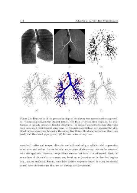
(a) (b) (c)<br />
(d) (e) (f)<br />
Figure 7.4: Illustration <strong>of</strong> the process<strong>in</strong>g steps <strong>of</strong> the airway tree reconstruction approach.<br />
(a) Volume render<strong>in</strong>g <strong>of</strong> the utilized dataset. (b) Tube detection filter response. (c) Centerl<strong>in</strong>es<br />
<strong>of</strong> <strong>in</strong>itially extracted tubular structures. (d) Initially extracted tubular structures<br />
with associated radii/tangent directions. (e) Group<strong>in</strong>g and l<strong>in</strong>kage step show<strong>in</strong>g the identified<br />
tubular structures belong<strong>in</strong>g the airway tree (blue), the discarded tubular structures<br />
(red), and the closed gaps (green). (f) Reconstructed airway tree.<br />
associated radius and tangent direction are <strong>in</strong>dicated us<strong>in</strong>g a cyl<strong>in</strong>der with appropriate<br />
orientation and radius. As can be seen, major parts <strong>of</strong> the airway tree can be extracted<br />
with this approach. However, two problems rema<strong>in</strong> that have to be addressed. First, the<br />
centerl<strong>in</strong>es <strong>of</strong> the tubular structures may break up at junctions or <strong>in</strong> disturbed regions<br />
(e.g., motion artifacts). Second, some false positive responses caused by other low density<br />
(dark) tube-like structures that are not airways are also present.


