Segmentation of 3D Tubular Tree Structures in Medical Images ...
Segmentation of 3D Tubular Tree Structures in Medical Images ...
Segmentation of 3D Tubular Tree Structures in Medical Images ...
Create successful ePaper yourself
Turn your PDF publications into a flip-book with our unique Google optimized e-Paper software.
5.3. Evaluation and Results 91<br />
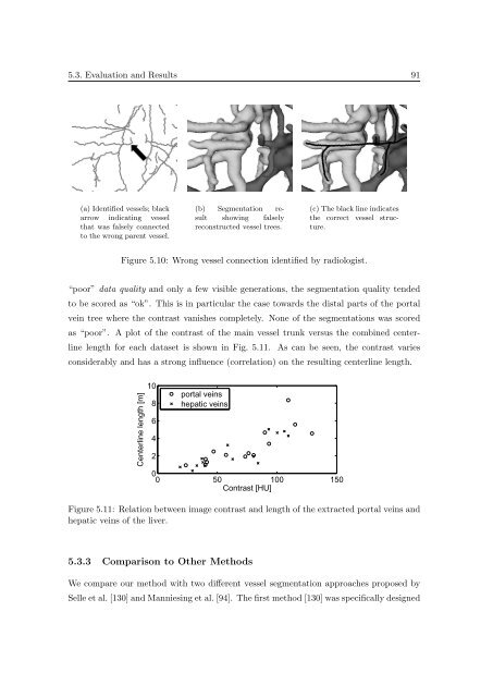
(a) Identified vessels; black<br />
arrow <strong>in</strong>dicat<strong>in</strong>g vessel<br />
that was falsely connected<br />
to the wrong parent vessel.<br />
(b) <strong>Segmentation</strong> result<br />
show<strong>in</strong>g falsely<br />
reconstructed vessel trees.<br />
(c) The black l<strong>in</strong>e <strong>in</strong>dicates<br />
the correct vessel structure.<br />
Figure 5.10: Wrong vessel connection identified by radiologist.<br />
“poor” data quality and only a few visible generations, the segmentation quality tended<br />
to be scored as “ok”. This is <strong>in</strong> particular the case towards the distal parts <strong>of</strong> the portal<br />
ve<strong>in</strong> tree where the contrast vanishes completely. None <strong>of</strong> the segmentations was scored<br />
as “poor”. A plot <strong>of</strong> the contrast <strong>of</strong> the ma<strong>in</strong> vessel trunk versus the comb<strong>in</strong>ed centerl<strong>in</strong>e<br />
length for each dataset is shown <strong>in</strong> Fig. 5.11. As can be seen, the contrast varies<br />
considerably and has a strong <strong>in</strong>fluence (correlation) on the result<strong>in</strong>g centerl<strong>in</strong>e length.<br />
Centerl<strong>in</strong>e length [m]<br />
10<br />
8<br />
6<br />
4<br />
2<br />
portal ve<strong>in</strong>s<br />
hepatic ve<strong>in</strong>s<br />
0<br />
0 50 100 150<br />
Contrast [HU]<br />
Figure 5.11: Relation between image contrast and length <strong>of</strong> the extracted portal ve<strong>in</strong>s and<br />
hepatic ve<strong>in</strong>s <strong>of</strong> the liver.<br />
5.3.3 Comparison to Other Methods<br />
We compare our method with two different vessel segmentation approaches proposed by<br />
Selle et al. [130] and Mannies<strong>in</strong>g et al. [94]. The first method [130] was specifically designed


