Segmentation of 3D Tubular Tree Structures in Medical Images ...
Segmentation of 3D Tubular Tree Structures in Medical Images ...
Segmentation of 3D Tubular Tree Structures in Medical Images ...
You also want an ePaper? Increase the reach of your titles
YUMPU automatically turns print PDFs into web optimized ePapers that Google loves.
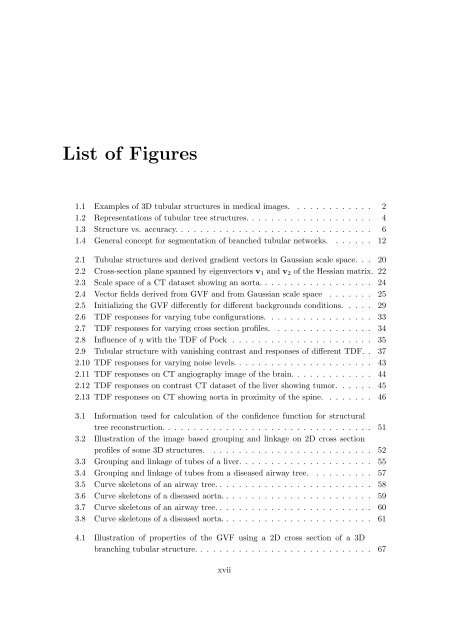
List <strong>of</strong> Figures<br />
1.1 Examples <strong>of</strong> <strong>3D</strong> tubular structures <strong>in</strong> medical images. . . . . . . . . . . . . 2<br />
1.2 Representations <strong>of</strong> tubular tree structures. . . . . . . . . . . . . . . . . . . . 4<br />
1.3 Structure vs. accuracy. . . . . . . . . . . . . . . . . . . . . . . . . . . . . . . 6<br />
1.4 General concept for segmentation <strong>of</strong> branched tubular networks. . . . . . . 12<br />
2.1 <strong>Tubular</strong> structures and derived gradient vectors <strong>in</strong> Gaussian scale space. . . 20<br />
2.2 Cross-section plane spanned by eigenvectors v 1 and v 2 <strong>of</strong> the Hessian matrix. 22<br />
2.3 Scale space <strong>of</strong> a CT dataset show<strong>in</strong>g an aorta. . . . . . . . . . . . . . . . . . 24<br />
2.4 Vector fields derived from GVF and from Gaussian scale space . . . . . . . 25<br />
2.5 Initializ<strong>in</strong>g the GVF differently for different backgrounds conditions. . . . . 29<br />
2.6 TDF responses for vary<strong>in</strong>g tube configurations. . . . . . . . . . . . . . . . . 33<br />
2.7 TDF responses for vary<strong>in</strong>g cross section pr<strong>of</strong>iles. . . . . . . . . . . . . . . . 34<br />
2.8 Influence <strong>of</strong> η with the TDF <strong>of</strong> Pock . . . . . . . . . . . . . . . . . . . . . . 35<br />
2.9 <strong>Tubular</strong> structure with vanish<strong>in</strong>g contrast and responses <strong>of</strong> different TDF. . 37<br />
2.10 TDF responses for vary<strong>in</strong>g noise levels. . . . . . . . . . . . . . . . . . . . . . 43<br />
2.11 TDF responses on CT angiography image <strong>of</strong> the bra<strong>in</strong>. . . . . . . . . . . . . 44<br />
2.12 TDF responses on contrast CT dataset <strong>of</strong> the liver show<strong>in</strong>g tumor. . . . . . 45<br />
2.13 TDF responses on CT show<strong>in</strong>g aorta <strong>in</strong> proximity <strong>of</strong> the sp<strong>in</strong>e. . . . . . . . 46<br />
3.1 Information used for calculation <strong>of</strong> the confidence function for structural<br />
tree reconstruction. . . . . . . . . . . . . . . . . . . . . . . . . . . . . . . . . 51<br />
3.2 Illustration <strong>of</strong> the image based group<strong>in</strong>g and l<strong>in</strong>kage on 2D cross section<br />
pr<strong>of</strong>iles <strong>of</strong> some <strong>3D</strong> structures. . . . . . . . . . . . . . . . . . . . . . . . . . 52<br />
3.3 Group<strong>in</strong>g and l<strong>in</strong>kage <strong>of</strong> tubes <strong>of</strong> a liver. . . . . . . . . . . . . . . . . . . . . 55<br />
3.4 Group<strong>in</strong>g and l<strong>in</strong>kage <strong>of</strong> tubes from a diseased airway tree. . . . . . . . . . 57<br />
3.5 Curve skeletons <strong>of</strong> an airway tree. . . . . . . . . . . . . . . . . . . . . . . . . 58<br />
3.6 Curve skeletons <strong>of</strong> a diseased aorta. . . . . . . . . . . . . . . . . . . . . . . . 59<br />
3.7 Curve skeletons <strong>of</strong> an airway tree. . . . . . . . . . . . . . . . . . . . . . . . . 60<br />
3.8 Curve skeletons <strong>of</strong> a diseased aorta. . . . . . . . . . . . . . . . . . . . . . . . 61<br />
4.1 Illustration <strong>of</strong> properties <strong>of</strong> the GVF us<strong>in</strong>g a 2D cross section <strong>of</strong> a <strong>3D</strong><br />
branch<strong>in</strong>g tubular structure. . . . . . . . . . . . . . . . . . . . . . . . . . . . 67<br />
xvii


