

Segmentation of 3D Tubular Tree Structures in Medical Images ...
Segmentation of 3D Tubular Tree Structures in Medical Images ...
Segmentation of 3D Tubular Tree Structures in Medical Images ...

You also want an ePaper? Increase the reach of your titles
YUMPU automatically turns print PDFs into web optimized ePapers that Google loves.
112 Chapter 7. Airway <strong>Tree</strong> <strong>Segmentation</strong><br />
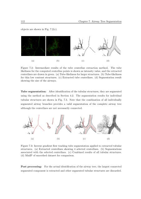
objects are shown <strong>in</strong> Fig. 7.2(c).<br />
(a) (b) (c) (d)<br />
Figure 7.2: Intermediate results <strong>of</strong> the tube centerl<strong>in</strong>e extraction method. The tube<br />
likel<strong>in</strong>ess for the computed centerl<strong>in</strong>e po<strong>in</strong>ts is shown as <strong>in</strong>tensity value, and the extracted<br />
centerl<strong>in</strong>es are drawn <strong>in</strong> green. (a) Tube-likel<strong>in</strong>ess for larger structures. (b) Tube-likel<strong>in</strong>ess<br />
for th<strong>in</strong> low contrast structures. (c) Extracted tube centerl<strong>in</strong>es. (d) <strong>Segmentation</strong> result<br />
show<strong>in</strong>g the size <strong>of</strong> the airways.<br />
Tube segmentation: After identification <strong>of</strong> the tubular structures, they are segmented<br />
us<strong>in</strong>g the method as described <strong>in</strong> Section 4.2. The segmentation results for <strong>in</strong>dividual<br />
tubular structures are shown <strong>in</strong> Fig. 7.3. Note that the comb<strong>in</strong>ation <strong>of</strong> all <strong>in</strong>dividually<br />
segmented airway branches provides a valid segmentation <strong>of</strong> the complete airway tree<br />
although the centerl<strong>in</strong>es are not necessarily connected.<br />
(a) (b) (c) (d)<br />
Figure 7.3: Inverse gradient flow track<strong>in</strong>g tube segmentation applied to extracted tubular<br />
structures. (a) Extracted centerl<strong>in</strong>es show<strong>in</strong>g 4 selected centerl<strong>in</strong>es. (b) <strong>Segmentation</strong>s<br />
associated with the selected centerl<strong>in</strong>es. (c) Comb<strong>in</strong>ed results <strong>of</strong> all tubular structures.<br />
(d) M<strong>in</strong>IP <strong>of</strong> smoothed dataset for comparison.<br />
Post process<strong>in</strong>g:<br />
For the actual identification <strong>of</strong> the airway tree, the largest connected<br />
segmented component is extracted and other segmented tubular structures are discarded.


