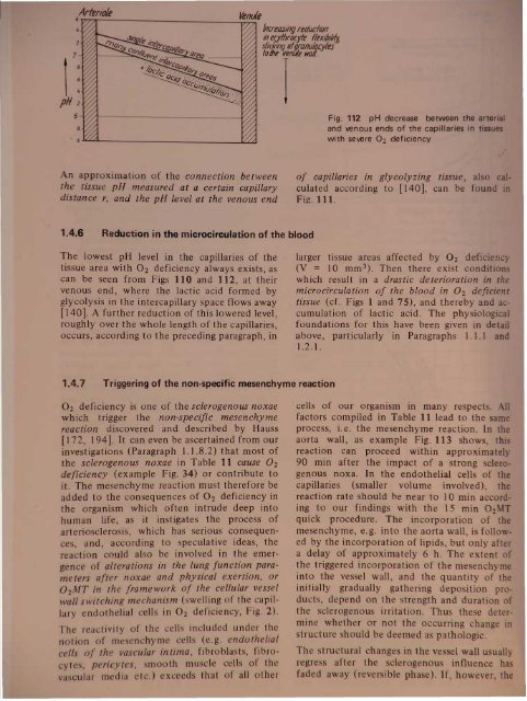
ventieFig. 112 pH decrease between the arteriand venous ends of the capillaries in tiwith severe 02 deficiencyAn approximation of the connection betweenthe tissue pH measured at a certain capillarydistance r, and the pH level at the venous endof capillaries in glycolyzing tissue, also calculated according to [140], can be found .Fig. Ill.1.4.6 Reduction in the microcirculation of the bloodThe lowest pH level in the capillaries of thetissue area with O 2 deficiency always exists, ascan be seen from Figs 110 and 112, at theirvenous end, where the lactic acid formed byglycolysis in the intercapillary space flows away[140]. A further reduction of this lowered level,roughly over the whole length of the capillaries,occurs, according to the preceding paragraph, inlarger tissue areas affected by O 2 deficien(V = 10 mm 3 ). Then there exist conditiowhich result in a drastic deterioration in themicrocirculation of the blood in O 2 deficienttissue (cf. Figs 1 and 75), and thereby and a cumulation of lactic acid. The physiologicalfoundations for this have been given in detailabove, particularly in Paragraphs 1.1.1 and1.2.1.1.4.7 Triggering of the non-specific mesenchyme reaction02 deficiency is one of the sclerogenous noxaewhich trigger the non-specific mesenchymereaction discovered and described by Hauss[] 72, ] 94]. It can even be ascertained from ourinvestigations (Paragraph 1.] .8.2) that most ofthe sclerogenous noxae in Table 11 cause O 2deficiency (example Fig. 34) or contribute toit. The mesenchyme reaction must therefore beadded to the consequences of O 2 deficiency inthe organism which often intrude deep intohuman life, as it instigates the process ofarteriosclerosis, which has serious consequences,and, according to speculative ideas, thereaction co ld also be involved in the emergenceof ~lterations in the lung function parametersafter noxae and physical exertion, or02MT in the framework of the cellular vesselwall switching mechanism (swelling of the capillaryendothelial cells in O 2 deficiency, Fig. 2).The reactivity of the cells included under thenotion of me enchyme cells (e.g. endothelialcelis of the vascular intima, fibroblasts, fibrocyte, pericytes, smooth mu de cell of thva cular m dia etc.) exceed that of all oth r. cells of our organism in many respects.factors compiled in Table 11 lead to the amprocess, i. e. the mesenchyme reaction. In thaorta wall, as example Fig. 113 shows t'reaction can proceed within approximatel90 min after the impact of a strong sclerogenousnoxa. In the endothelial cells of thcapillaries (smaller volume involved) threaction rate should be near to 10 min ac ortiingto our findings with the 15 min O 2quick procedure. The incorporation of thmesenchyme, e.g. into the aorta wall i folIo ed by the incorporation of lipid but onl afa delay of approximately 6 h. The t nt. the triggered incorporation of the me n h minto the vessel wall, and the quantit f thinitially gradually gath ring d p iti n pr·ducts, depend on the tr ngth and durati nthe cl rogenou irritation. Thu th d r·mine wh th r or not th urring h n intructur hould b d m d a path 1Th tm tural hang in th 1 11 u u 11r gr aft r th 1 r nou influ nfad d away (r v ibl ph' . If howl~ve
of clerogenou noxae. 1 Di turbing factors which, according to W.H. Hauss, regularly trigger thenchyme reaction (172), detected by the increased rate of 3SS-sulfate incorporation into the sulfo-lPOly accharide of the ground substance of connective tissue in various organs (animal experiments)I of oxygen 2h pOtensiony rtension (Fig. 39)rationsinsi factionsnicotineforeign proteinallergic reactionso foreign substancesover4xertion2 mechanical stress13 interbrein stimulationemotional stimuli6 noise6 hormones17 influences of dietDisturbing factorshypobaric chamber with P0 2values < 150 mmHgblood pressure amplitude too low (e.g. < 25 mmHg)reduced kidney circulation; hypertension (systol. blood pressure> 200 mmHg)anesthesia etc.endotoxins, staphylococcus toxin, diphtheria toxinpasteurella multocida, staphylococciinhalation of cigarette smokealbumin injectionserum shock, Arthus' phenomenoncroton oil, plastic productsexcessive movement in the treadmilllaceration of muscle, dilatation of the aortathalamus stimulation by means of electric.currentbinding, restricted movementroaring, uniform random noiseadrenaline, testosterone, thyroxine, cortisone, glucagonsclerogenous diets1 Most of these noxae deteriorate the oxygen status2 Subnormal values of Po or '11; locally reduced 02 delivery to the vessel wall; mechanical irritation at ramificationsin the arterial systir:t rtsclerogenous irritation or O 2 deficiency lastsfor a long time (reduced clearance capacity dueto energy deficiency) and particularly if suchong-lasting conditions frequently recur, thenthe non-decomposable deposition productsfrom the metabolism (collagen fibre bundles,calcification etc.) accumulate in the way shownby the electron micrographs in [I 72]. Thedamage to the vessel wall caused by thesebreakdown products no longer recedes (irreversiblephase), as the increase of the diffusionength in both directions of the vessel wallradial, central) begins definitively to limit theetabolism there. A final stabilization of theypertrophic structure thus oc~urs. We ther~forebelieve that the pathologIcal changes Intructure of the vessel wall remain reversible fora long time. The important th~ng is. that thearteriosclerosis be combatted In thIS phase,fore the irreversible wall degeneration finallyes place. We have already seen [2, 8, 106]e reversal of the sclerogenous noxa "oxygenficiency" as a method of renormalization of'he velieI wall structure in the reversible phase,the 02MT in a highly intensive variant. ted to the part of the body in question.ct f ting discussed in Paragraph 5.3.9Ie 42) hould al 0 be mentioned as aIUf'tl'llfll!r method for the renormalization oftt8Jiosci rotically degenerated wall of arterieand arterioles, and of degenerated layers of thealveocapillary system of the lung.According to [172] the pathological changes inthe vessel wall structure caused by the nonspecificmesenchyme reaction affect mainlarterial vessels and their ramifications. Despitethis, this reaction could always re ult in adeterioration of the O 2 supply to the ti sue viacapillaries, above a certain intensity. Thideterioration should result from the reductionof the blood flow (cross-section narrowingreduction in vasomotoric adaptability and inthe air chamber effect) in sclerotically degeneratedarteries.A related reaction in the fine blood capillariecauses the vessel wall thickening di cos ed in1.1.1, and thereby a narrowing of the lumenand even a direct hampering of oxygen and ubstratediffusion to the intercapillar pa . Thbasal membrane thickening of the capillori sdue to protein depo its i a w ll-kno n ph nomenonin diabetic (mi ro)angi pathi andkidney di ea e. Th me pro an Ioccur in th m mbran f th m lin h(Paragraph 1.4.11) and th n I d t th n ur pathie of th diab ti . In nn ti nih hiwe hould mention that th patho ni b Imembran thi k nin n b br n dtri t fa ting ( nd I
- Page 1 and 2:
sand functl ni ove d cellular capil
- Page 3:
8 1. Physiological fou~dationsmmHga
- Page 6 and 7:
Basic mechanisms and functions 11e
- Page 8 and 9:
Basic mechanisms and functions 13pr
- Page 11 and 12:
500r-T_-+5__r-_-+1O__,....-_~15.1O-
- Page 13 and 14:
~IUI1!J ~atpWorMs---~~·tIIlDkeJ \I
- Page 15 and 16:
y r na onabS,olu'te clla.ra~t ri ti
- Page 17 and 18:
100907065"I 4y iological foundation
- Page 19 and 20:
1. hy lologlcal foundations70"''''J
- Page 21 and 22:
CII'Cli8LC outl)ut VAr;lllil i e re
- Page 23 and 24:
-'0 y 101091cai oundatlons80%6050l~
- Page 25 and 26:
,..._........,.--~"'!""!!'!~~----_.
- Page 27 and 28:
hi v d in h arrangement shown above
- Page 29 and 30:
34 1. Physiological foundations70%6
- Page 31 and 32:
logical foundationsu .on in the opt
- Page 33 and 34:
1. Physiological foundations. ~ la~
- Page 35 and 36:
II foundationsI tem IBPtrsons with
- Page 37:
1. Physiological foundationsAirdosi
- Page 40 and 41:
110N.ttJJOt~1. Physiological founda
- Page 42 and 43:
1. Phy iological foundationsAnoO;HT
- Page 44 and 45:
hysiological foundations...fJJHT(JI
- Page 46 and 47:
1. Physiological foundationsCOCOM I
- Page 48 and 49:
g r d, whi h con iderably contribut
- Page 50 and 51:
logical foundations13IrPtz12.......
- Page 52 and 53:
,. Physiological foundationsIf-RoN
- Page 54 and 55:
1. Physiological foundationsAll"i1t
- Page 56 and 57: ~~itItINitIIJq/s N 31 54 52~IBIP~-.
- Page 58 and 59: BDuring OzMTprocedurecAfter OzMTpro
- Page 60 and 61: A Bi3lrfss 1'1oafofmovement 8 0pl'r
- Page 62 and 63: Table 6 PO b measurements of unsele
- Page 64 and 65: serert~ hyperoxia pure 0 } .distres
- Page 66 and 67: IIstress qirI/iII mn wryhJI1IJSISLo
- Page 68 and 69: A/JtfJtnk«P~1Mtrr'ttriIWIItJfIS~.~
- Page 70 and 71: Table 7 The change in the architect
- Page 72 and 73: nsMslor rotd'1uI.-Jl!Jg1"/11(J(Jrfr
- Page 74 and 75: IonsFig. 74 Histological pictures s
- Page 76 and 77: IonseM' Selecfofherm equipment-_s1S
- Page 78 and 79: .....-------................',..,-n
- Page 80 and 81: eM/rot gro"/!.Histologicallyconfirm
- Page 82 and 83: A1er tnit,almonipulotionsand III«l
- Page 84 and 85: Q'CSlNltftning1A. QfQ/-fJlf1inguQ
- Page 86 and 87: otllm'lv.2 h th ju tifi d pro p ct
- Page 88 and 89: t48xlD J WsEhmmahooofMe ~crdieal, c
- Page 90 and 91: old°z-/ImitMW mode of calcu/aflonC
- Page 92 and 93: he cylindr'cal area upplied by the'
- Page 94 and 95: Tn-T-~-----1~--~--~JaluralttJtranSi
- Page 96 and 97: aPI)Ii:lcati?n with a patially very
- Page 98 and 99: are triggered. At the same site, ho
- Page 100 and 101: n respired again, the drop in respi
- Page 102 and 103: Exholationtubeloop aroundcmebroncll
- Page 104 and 105: [Xeftt of,t"". Respiratorv... stand
- Page 108 and 109: BIncorporation ofJ5s-slI"ate intofh
- Page 110 and 111: ~LFig. 115 Electron micrograph of h
- Page 112 and 113: 12111098V ~ .-"~"' .....b""", ".1Tt
- Page 114 and 115: ,,,;tt cola;, ofH1e 88JIJitKIJIIl "
- Page 116 and 117: "-1r-180 I'\112007(J)Jr' '- jwry¥I