Chapter-1 / Physiological Foundations - WHNLive Public Library
Chapter-1 / Physiological Foundations - WHNLive Public Library
Chapter-1 / Physiological Foundations - WHNLive Public Library

Create successful ePaper yourself
Turn your PDF publications into a flip-book with our unique Google optimized e-Paper software.
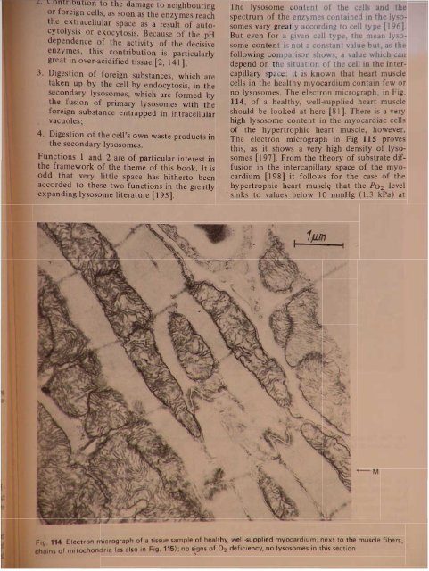
ntlu Ion to the damage to neighbouringr foreign cells, a 000 a the enzyme reachth extracellular space a a result of autoytolyi or exocyto is. Because of the pHdependence of the activity of the decisivenzy~e, this contribution is particularlygr at In over-acidified tissue [2, 141];Dige tion of foreign substances, which aretaken up by the cell by endocytosis, in thesecondary lysosomes, which are formed bythe fusion of primary lysosomes with theforeign substance entrapped in intracellularvacuoles;4. Digestion of the cell's own waste products inthe secondary lysosomes.Functions 1 and 2 are of particular interest inthe framework of the theme of this book. It isodd that very little space has hitherto beenaccorded to these two functions in the greatlyexpanding lysosome literature [195].The lysosome content of the cells and thespectrum of the enzymes contained in the ly 0somes vary greatly according to ce)) type [196].But even for a given cell type, the mean lysosomecontent is not a constant value but, as thefollowing comparison shows, a value which candepend on the situation of the cell in the intercapillaryspace: it is known that heart musclecells in the healthy myocardium contain few orno lysosomes. The electron micrograph, in Fig.114, of a healthy, well-supplied heart muscleshould be looked at here [81]. There is a veryhigh lysosome content in the myocardiac cellsof the hypertrophic heart muscle, however.The electron micrograph in Fig. 115 provesthis, as it shows a very high density of lysosomes(197]. From the theory of substrate diffusionin the intercapillary space of the myocardium[198] it follows for the case of thehypertrophic heart muscl~ that the P02 level. sinks to values below 10 mmHg (1.3 kPa) at1pmJ-........----t~MIg.114 Electron micrograph of a tissue sample .of healthy, vve~l~uPPlied myocardiu~; n~xt to .the muscle fibers,inS of mitochondria (as also in Fig. 115); no I~gns of 02 deficiency, no Iysosomes In thl section