Download the ESMO 2012 Abstract Book - Oxford Journals
Download the ESMO 2012 Abstract Book - Oxford Journals
Download the ESMO 2012 Abstract Book - Oxford Journals
You also want an ePaper? Increase the reach of your titles
YUMPU automatically turns print PDFs into web optimized ePapers that Google loves.
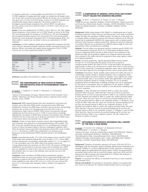
or cutaneous melanoma. A survival update was performed on 31 March 2011.<br />
CS-PHP melphalan 3.0 mg/kg ideal body weight was infused into <strong>the</strong> hepatic artery<br />
over 30 min with concurrent extracorporeal filtration for 60 min. Up to 6 treatments<br />
were given every 4–8 wks. In <strong>the</strong> BAC group, crossover to CS-PHP melphalan was<br />
permitted after hepatic disease progression. The primary endpoint was<br />
investigator-assessed hepatic progression-free survival (hPFS). An exploratory<br />
post-hoc analysis of pts who crossed from BAC to CS-PHP vs. BAC-only pts was<br />
also performed.<br />
Results: 93 pts were randomized to CS-PHP (n = 44) or BAC (n = 49). After hepatic<br />
disease progression, 28 pts crossed over to CS-PHP. Results are shown in <strong>the</strong> Table.<br />
The most common grade 3/4 toxicities in CS-PHP pts (n = 40) were hematological<br />
peri-procedural thrombocytopenia (73%) and anemia (55%) and post-procedural<br />
(beyond day 4 post-treatment) neutropenia (93%) or thrombocytopenia (83%). The<br />
safety profile in crossover pts was similar to that in pts randomized to CS-PHP<br />
melphalan.<br />
Conclusions: CS-PHP melphalan significantly prolonged hPFS compared with BAC<br />
in pts with liver-dominant metastatic melanoma, <strong>the</strong>reby meeting <strong>the</strong> primary study<br />
objective. Efficacy was similar after hepatic disease progression in BAC-CS-PHP<br />
crossover pts as in those randomized initially to CS-PHP.<br />
Median<br />
hPFS, Hazard ratio Median Hazard ratio<br />
Treatment group n mo (95% CI) OS, mo (95% CI)<br />
CS-PHP 44 8.0 0.35 (0.23-0.54) 9.8 1.08 (0.69-1.68)<br />
BAC 49 1.6 P < 0.0001 9.9 NS<br />
BAC only 21 1.6 0.32 4.1 0.33<br />
BAC →<br />
CS-PHP crossover<br />
28 8.8 15.3<br />
Disclosure: All authors have declared no conflicts of interest.<br />
1142P THE CONCORDANCE OF HER2 STATUS IN PRIMARY<br />
AND METASTATIC SITES OF EXTRAMAMMARY PAGET’S<br />
DISEASE<br />
R. Tanaka 1 , Y. Sasajima 2 , H. Tsuda 2 , K. Namikawa 1 , A. Tsutsumida 1 ,<br />
N. Yamazaki 1<br />
1 Division of Dermatologic Oncology, National Cancer Center Hospital, Tokyo,<br />
JAPAN, 2 Pathology and Clinical Laboratory Division, National Cancer Center<br />
Hospital, Tokyo, JAPAN<br />
Background: HER2-targeted <strong>the</strong>rapies have been introduced to treat breast and<br />
stomach cancers that show HER2 protein overexpression and/or HER2 gene<br />
amplification. However, <strong>the</strong> HER2 status of primary tumors and <strong>the</strong>ir corresponding<br />
metastatic sites is shown to be heterogeneous in less than 20% of cases. In<br />
extramammary Paget’s disease (EMPD), HER2 protein overexpression and gene<br />
amplification has been shown to occur in 5-80% of cases; however, knowledge<br />
regarding <strong>the</strong> concordance of HER2 status in primary and metastatic sites of EMPD<br />
is limited. The aim of this study was to clarify <strong>the</strong> concordance rate of HER2 status<br />
in <strong>the</strong> primary tumors and metastatic sites of EMPD.<br />
Methods: Twenty-six tissue blocks of primary tumors and corresponding lymph<br />
node metastases were subjected to an immunohistochemistry (IHC) analysis. The<br />
IHC scores were classified into four groups (0 to 3+) according to <strong>the</strong> American<br />
Society of Clinical Oncology/College of American Pathologists Guidelines (2007).<br />
When <strong>the</strong> primary tumors and lymph node metastases showed IHC scores of ei<strong>the</strong>r<br />
2+ or 3 + , <strong>the</strong> presence of HER2 gene amplification was examined using<br />
fluorescence in situ hybridization (FISH) and dual-colored in situ hybridization<br />
(DISH).<br />
Results: mmunohistochemically, 27% (7/26) of <strong>the</strong> primary tumors and 38% (10/26)<br />
of <strong>the</strong> lymph node metastases showed IHC scores of ei<strong>the</strong>r 2+ or 3+. When HER2<br />
protein overexpression was classified as being ei<strong>the</strong>r positive (2 + , 3+) or negative (0,<br />
1+), <strong>the</strong> concordance rate of <strong>the</strong> HER2 status of <strong>the</strong> primary tumors and<br />
corresponding lymph node metastases was 85% (22/26). The presence of HER2 gene<br />
amplification was examined in six primary tumors and 10 metastatic sites. HER2<br />
gene was amplified in a total of seven cases (27%): four cases in both <strong>the</strong> primary<br />
and metastatic sites, two cases in <strong>the</strong> metastatic sites only and one case in <strong>the</strong><br />
primary site only.<br />
Conclusion: Good concordance of HER2 protein overexpression and gene<br />
amplification status was seen in <strong>the</strong> primary tumors and corresponding lymph node<br />
metastases of patients with EMPD. Fur<strong>the</strong>rmore, <strong>the</strong> HER2 gene was found to be<br />
always amplified in cases with an IHC score of 3+ and in two cases with an IHC<br />
score of 2+ in <strong>the</strong> lymph node metastases, which is suitable to be targeted for<br />
<strong>the</strong>rapy.<br />
Disclosure: All authors have declared no conflicts of interest.<br />
Annals of Oncology<br />
1143P A COMPARISON OF GENERAL POPULATION AND PATIENT<br />
UTILITY VALUES FOR ADVANCED MELANOMA<br />
A. Batty 1 , B. Winn 1 , L. Pericleous 2 ,D.Rowen 3 , D. Lee 1 , T. Nikoglou 2<br />
1 Health Economics, BresMed, Sheffield, UNITED KINGDOM, 2 Health Economics<br />
and Outcomes, Bristol Myers Squibb, Uxbridge, UNITED KINGDOM, 3 School of<br />
Health and Related Research, University of Sheffield, Sheffield, UNITED<br />
KINGDOM<br />
Background: Health-related quality of life (HRQL) is a fundamental part of health<br />
technology assessment. Utility values are vital because <strong>the</strong>y can be used as preference<br />
weights and allow <strong>the</strong> calculation of HRQL benefits. The objective of this study was<br />
to compare utilities calculated for patients with advanced melanoma in <strong>the</strong> Phase III<br />
clinical trial for ipilimumab (MDX010-20) using a generic and a condition-specific<br />
preference-based measure to utilities produced by vignettes for advanced melanoma.<br />
A secondary objective was to determine how different analyses might be used most<br />
appropriately within cost-effectiveness modelling.<br />
Methods: The trial utilities were generated using <strong>the</strong> condition-specific EORTC-8D<br />
(1,190 observations) and generic SF-6D (1,157 observations) preference-based<br />
measures. Progression-status and time-to-death analyses were conducted on <strong>the</strong><br />
patient-level data and <strong>the</strong> predictive abilities were compared. Patient-level results<br />
were compared to <strong>the</strong> utilities derived for progression status using vignettes valued<br />
by <strong>the</strong> general population.<br />
Results: On disease progression, vignette-generated utilities showed a greater<br />
decrease (0.77 to 0.59) than ei<strong>the</strong>r <strong>the</strong> generic SF-6D (0.64 to 0.619) or<br />
condition-specific EORTC-8D (0.801 to 0.763). SF-6D and EORTC-8D showed a<br />
large decrease in utility in <strong>the</strong> 180 days prior to death (from 0.826 to 0.628 and from<br />
0.655 to 0.505, respectively). Compared to progression status, time to death showed a<br />
lower Root Mean Squared Error and higher R2 when used to predict patient utility.<br />
Conclusion: Practitioners should carefully analyse patient level utility data prior to<br />
constructing economic models as standard measures, such as progression status,<br />
may not fully capture <strong>the</strong> patient experience. Similarly, where vignettes are valued<br />
to represent health states in an economic model, <strong>the</strong>ir applicability to <strong>the</strong> disease<br />
and patient population should be carefully scrutinised. The use of standard<br />
progression based cost-effectiveness modelling techniques may not be appropriate<br />
for this disease. Consequently, <strong>the</strong>re are implications for <strong>the</strong> analysis of utility<br />
information in future studies and <strong>the</strong> methods of cost-effectiveness modelling used<br />
for cancer treatments.<br />
Disclosure: A. Batty: BresMed were funded by BMS to conduct <strong>the</strong> analysis<br />
presented within this paper. O<strong>the</strong>r than this I have not received any personal<br />
funding and do not have any personal interest in BMS or any competitor products.<br />
B. Winn: BresMed were funded by BMS to conduct <strong>the</strong> analysis presented within<br />
this paper. O<strong>the</strong>r than this I have not received any personal funding and do not<br />
have any personal interest in BMS or any competitor products. L. Pericleous: I have<br />
worked for BMS. O<strong>the</strong>r than this I have not received any personal funding and do<br />
not have any personal interest in BMS or any competitor products. D. Lee:<br />
BresMed were funded by BMS to conduct <strong>the</strong> analysis presented within this paper.<br />
O<strong>the</strong>r than this I have not received any personal funding and do not have any<br />
personal interest in BMS or any competitor products. T. Nikoglou: I work for BMS.<br />
O<strong>the</strong>r than this I have not received any personal funding and do not have any<br />
personal interest in BMS or any competitor products. All o<strong>the</strong>r authors have<br />
declared no conflicts of interest.<br />
1144 CETUXIMAB IN METASTATIC SQUAMOUS CELL CANCER<br />
OF THE SKIN: A CASE SERIES<br />
K. Conen 1 , N. Fischer 2 , G.F. Hofbauer 3 , B. Shafaeddin-Schreve 3 ,<br />
R. Win<strong>the</strong>rhalder 4 , C. Rochlitz 5 , A. Zippelius 1<br />
1 Medical Oncology, University Hospital Basel, Basel, SWITZERLAND, 2 Medical<br />
Oncology, Kantonsspital Winterthur, Winterthur, SWITZERLAND,<br />
3 Dermatologische Klinik, Universitätsspital Zürich, Zürich, SWITZERLAND,<br />
4 Medical Oncology, Kantonsspital Luzern, Luzern, SWITZERLAND, 5 Medical<br />
Onkology, University Hospital Basel, Basel, SWITZERLAND<br />
Background: Treatment of metastatic squamous cell cancer of <strong>the</strong> skin (SCCS) is<br />
challenging. Only few <strong>the</strong>rapeutic options including cisplatin-based combinations are<br />
currently available. In a recently published phase II trial, cetuximab, a monoclonal<br />
antibody against <strong>the</strong> epidermal growth factor receptor (EGRF), has demonstrated a<br />
promising clinical activity with an overall 69% disease control rate (DCR) and 28%<br />
response rate (RR) in patients with unresectable SCCS. Whe<strong>the</strong>r this treatment is<br />
also effective in metastatic SCCS is unclear.<br />
Purpose: To summarize <strong>the</strong> efficacy of cetuximab at any line in a series of patients<br />
with metastatic SCCS.<br />
Patients and methods: We performed a retrospective analysis of six patients from<br />
four centres in Switzerland. We collected standard baseline data by reviewing <strong>the</strong><br />
ix372 | <strong>Abstract</strong>s Volume 23 | Supplement 9 | September <strong>2012</strong>


