als PDF - AGA
als PDF - AGA
als PDF - AGA
Erfolgreiche ePaper selbst erstellen
Machen Sie aus Ihren PDF Publikationen ein blätterbares Flipbook mit unserer einzigartigen Google optimierten e-Paper Software.
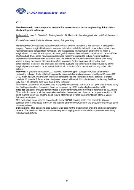
27. <strong>AGA</strong>-Kongress 2010 - Wien<br />
V 11<br />
New biomimetic nano-composite material for osteochondral tissue engineering: Pilot clinical<br />
study at 3 years follow-up<br />
Zaffagnini S., Kon E., Filardo G., Delcogliano M., Di Martino A., Marcheggiani Muccioli G.M., Marcacci<br />
M.<br />
Rizzoli Orthopaedic Institute, Biomechanics, Bologna, Italy<br />
Introduction: Chondral and osteochondral articular defects represent a key concern in orthopedic<br />
surgery. Current surgical techniques to repair osteochondral defects lead to poor subchondral bone<br />
regeneration and fibrocartilage formation, often associated with joint pain and stiffness. From a<br />
surgical and commercial standpoint, an ideal graft for osteochondral defect repair would be an off-theshelf<br />
product; thus, some new biomateri<strong>als</strong> were recently proposed to induce “in situ” cartilage<br />
regeneration after direct transplantation onto the defect site.We performed this clinical pilot study<br />
where a newly developed biomimetic scaffold was used for the treatment of chondral and<br />
osteochondral lesions of the knee joint in order to evaluate the safety and the reproducibility of the<br />
surgical procedure and in order to test the intrinsic potential of the device without any other cells<br />
culture.<br />
Methods: A gradient composite O.C. scaffold, based on type-I collagen-HA, was obtained by<br />
nucleating collagen fibrils with hydroxyapatite nanoparticles at physiological conditions 30 cases (9F,<br />
21M, mean age 29,3 years) with knee osteochondral lesions (8 medial femoral condyle, 5 lateral<br />
condyle, 12 patella, 8 femoral trochlea) were treated with scaffold implantation from January 2007 to<br />
July 2007. The lesions size went from 2 cm2 to 6 cm2.<br />
The clinical outcome of all patients was analyzed prospectively, at 6 moths at 1 year and 2 years using<br />
the Cartilage standard Evaluation Form as proposed by ICRS and an high resolution MRI .<br />
Results: Statistical analysis demonstrated a significant improvement from pre-operative to 12, 24 and<br />
36 months follow up in all the parameter evaluated. Moreover, we observed an improvement from 12<br />
to 24 months follow-up, and the good results obtained at 2 years were maintained at the 3 years<br />
follow-up evaluation.<br />
MRI evaluation was analyzed according to the MOCART scoring scale. The complete filling of<br />
cartilage defect was noted in 80% of the patients and the congruency of the articular surface was seen<br />
in same patients.<br />
Conclusions: This open one-step surgery was used for the treatment of chondral and osteochondral<br />
defects. The results of this technique are very encouraging and show satisfactory results even in big<br />
ostechondral defects.<br />
11


