- Page 1 and 2:
The Australian Immunisation Handboo
- Page 3 and 4:
FOREWORD Since 1932, when Governmen
- Page 5 and 6:
TABLE OF CONTENTS PART 1 INTRODUCTI
- Page 7 and 8:
LIST OF TABLES Table 2.1.1: Pre-vac
- Page 9 and 10:
List 4.13.1: Conditions associated
- Page 11 and 12:
PREFACE The 10th edition of The Aus
- Page 13 and 14:
Secretariat support, Australian Tec
- Page 15 and 16:
PART 1 INTRODUCTION TO THE AUSTRALI
- Page 17 and 18:
1.2 DEVELOPMENT OF THE 10TH EDITION
- Page 19 and 20:
1.3 HOW TO USE THE 10TH EDITION HAN
- Page 21 and 22:
1.4 WHAT’S NEW All chapters have
- Page 23 and 24:
2.2 Administration of vaccines •
- Page 25 and 26:
• The section on vaccination of p
- Page 27 and 28:
4.6 Human papillomavirus • HPV va
- Page 29 and 30:
• For Aboriginal and Torres Strai
- Page 31 and 32:
Part 5 Passive immunisation • Inf
- Page 33 and 34:
without the harmful consequences of
- Page 35 and 36:
(vaccine failure). Often such infec
- Page 37 and 38:
will occur following receipt of a s
- Page 39 and 40:
cases, both the doctor issuing the
- Page 41 and 42:
Should a child or adolescent refuse
- Page 43 and 44:
• check that the correct time int
- Page 45 and 46:
Note: Please discuss this informati
- Page 47 and 48:
Condition or circumstance of person
- Page 49 and 50:
Condition or circumstance of person
- Page 51 and 52:
Table 2.1.3: Live attenuated parent
- Page 53 and 54:
An online ‘catch-up calculator’
- Page 55 and 56:
Use of serological testing to guide
- Page 57 and 58:
• For some vaccines, catch-up vac
- Page 59 and 60:
Figure 2.1.1: Catch-up worksheet fo
- Page 61 and 62:
Vaccine Minimum age for 1st dose in
- Page 63 and 64:
Table 2.1.6: Number of vaccine dose
- Page 65 and 66:
Catch-up guidelines for individual
- Page 67 and 68:
If 13vPCV is not available, and 10v
- Page 69 and 70:
Previous vaccination history 2 prev
- Page 71 and 72:
PART 2 VACCINATION PROCEDURES 57 Ta
- Page 73 and 74:
PART 2 VACCINATION PROCEDURES 59 Ta
- Page 75 and 76:
Catch-up schedules for persons ≥1
- Page 77 and 78:
For additional details on these rec
- Page 79 and 80:
2.2 ADMINISTRATION OF VACCINES 2.2.
- Page 81 and 82:
• Never freeze a vaccine after it
- Page 83 and 84:
PART 2 VACCINATION PROCEDURES 69 In
- Page 85 and 86:
2.2.5 Vaccine injection techniques
- Page 87 and 88:
Interruption to a vaccination If th
- Page 89 and 90:
2.2.7 Positioning for vaccination I
- Page 91 and 92:
Children ≥12 months of age Cuddle
- Page 93 and 94:
2.2.8 Identifying the injection sit
- Page 95 and 96:
• Place the palm over the greater
- Page 97 and 98:
2.2.9 Administering multiple vaccin
- Page 99 and 100:
2.3 POST-VACCINATION 2.3.1 Immediat
- Page 101 and 102:
Management of an immediate adverse
- Page 103 and 104:
Management of anaphylaxis Rapid IM
- Page 105 and 106:
Autoinjectors are generally not app
- Page 107 and 108:
Australia. 16 This vaccine is no lo
- Page 109 and 110:
Any serious or unexpected adverse e
- Page 111 and 112:
Consumers and immunisation service
- Page 113 and 114:
When relevant, immunisation service
- Page 115 and 116:
National Human Papillomavirus Vacci
- Page 117 and 118:
Immunisation service providers enro
- Page 119 and 120:
PART 3 VACCINATION FOR SPECIAL RISK
- Page 121 and 122:
Thus, a vaccine to prevent Hib dise
- Page 123 and 124:
3.1.2 Adults Hepatitis B Indigenous
- Page 125 and 126:
een low in younger Indigenous adult
- Page 127 and 128:
3.2 VACCINATION FOR INTERNATIONAL T
- Page 129 and 130:
• vaccination history (including
- Page 131 and 132:
departure to allow for the period w
- Page 133 and 134:
Selected vaccines based on travel i
- Page 135 and 136:
Tick-borne encephalitis Tick-borne
- Page 137 and 138:
PART 3 VACCINATION FOR SPECIAL RISK
- Page 139 and 140:
PART 3 VACCINATION FOR SPECIAL RISK
- Page 141 and 142:
3.2.5 Vaccinating the traveller wit
- Page 143 and 144:
• Travel health and quarantine se
- Page 145 and 146:
whether the AEFI is likely to recur
- Page 147 and 148:
(see Appendix 1 Contact details for
- Page 149 and 150:
PART 3 VACCINATION FOR SPECIAL RISK
- Page 151 and 152:
PART 3 VACCINATION FOR SPECIAL RISK
- Page 153 and 154:
PART 3 VACCINATION FOR SPECIAL RISK
- Page 155 and 156:
PART 3 VACCINATION FOR SPECIAL RISK
- Page 157 and 158:
avoided, except in situations where
- Page 159 and 160:
3.3.3 Vaccination of immunocompromi
- Page 161 and 162:
Use of live viral or live bacterial
- Page 163 and 164:
Influenza vaccination is recommende
- Page 165 and 166:
PART 3 VACCINATION FOR SPECIAL RISK
- Page 167 and 168:
PART 3 VACCINATION FOR SPECIAL RISK
- Page 169 and 170:
Haematopoietic stem cell transplant
- Page 171 and 172:
Vaccine Months after HSCT Comments
- Page 173 and 174:
depending on the number of vaccines
- Page 175 and 176:
Persons with functional or anatomic
- Page 177 and 178:
Table 3.3.5: Recommendations for va
- Page 179 and 180:
Persons with autoimmune diseases an
- Page 181 and 182:
Table 3.3.6: Recommended intervals
- Page 183 and 184:
3.3.7 Vaccination of persons at occ
- Page 185 and 186:
Occupation Vaccine Providers of hom
- Page 187 and 188:
against certain vaccine-preventable
- Page 189 and 190:
3.3.11 Vaccination of persons who i
- Page 191 and 192:
waters. All cases of cholera report
- Page 193 and 194:
Children aged 2-6 years Three doses
- Page 195 and 196:
4.1.11 Adverse events The inactivat
- Page 197 and 198:
4.2.4 Vaccines Diphtheria toxoid is
- Page 199 and 200:
• Adacel - Sanofi Pasteur Pty Ltd
- Page 201 and 202:
antibodies at an age when waning of
- Page 203 and 204:
children aged
- Page 205 and 206:
4.3 HAEMOPHILUS INFLUENZAE TYPE B 4
- Page 207 and 208:
• Hiberix - GlaxoSmithKline (PRP-
- Page 209 and 210:
4.3.7 Recommendations Infants A Hib
- Page 211 and 212:
4.3.11 Public health management of
- Page 213 and 214:
In recent years, hepatitis A notifi
- Page 215 and 216:
Inactivated hepatitis A vaccines ar
- Page 217 and 218:
Co-administration with other vaccin
- Page 219 and 220:
Recommendations for the use of comb
- Page 221 and 222:
4.4.10 Adverse events The most comm
- Page 223 and 224:
4.5.3 Epidemiology The prevalence o
- Page 225 and 226:
4.5.4 Vaccines Monovalent hepatitis
- Page 227 and 228:
For older children and young adults
- Page 229 and 230:
Vaccine Age of vaccine recipient Do
- Page 231 and 232:
Combination hepatitis A/hepatitis B
- Page 233 and 234:
Management of infants born to mothe
- Page 235 and 236:
Household or other close (household
- Page 237 and 238:
with occult hepatitis B infection.
- Page 239 and 240:
immune memory persists and is thoug
- Page 241 and 242:
to suggest that a higher proportion
- Page 243 and 244:
Table 4.5.3: Post-exposure prophyla
- Page 245 and 246:
4.6 HUMAN PAPILLOMAVIRUS 4.6.1 Viro
- Page 247 and 248:
women. The prevalence of high-risk
- Page 249 and 250:
• Gardasil - CSL Limited/Merck &
- Page 251 and 252:
If scheduled doses have been missed
- Page 253 and 254:
However, some adult males may gain
- Page 255 and 256:
4.6.9 Contraindications The only ab
- Page 257 and 258:
4.7 INFLUENZA 4.7.1 Virology The in
- Page 259 and 260:
Figure 4.7.1: Influenza notificatio
- Page 261 and 262:
Always check annual seasonal influe
- Page 263 and 264:
7.5 µg of viral haemagglutinin (in
- Page 265 and 266:
Table 4.7.1: Recommended doses of i
- Page 267 and 268:
• Chronic respiratory conditions,
- Page 269 and 270:
Residents of residential aged care
- Page 271 and 272:
influenza vaccine prior to administ
- Page 273 and 274:
4.8 JAPANESE ENCEPHALITIS 4.8.1 Vir
- Page 275 and 276:
28 days following vaccination with
- Page 277 and 278:
When using JEspect in children aged
- Page 279 and 280:
4.8.8 Pregnancy and breastfeeding I
- Page 281 and 282:
4.9 MEASLES 4.9.1 Virology Measles
- Page 283 and 284:
4.9.4 Vaccines Monovalent measles v
- Page 285 and 286:
4.9.6 Dosage and administration The
- Page 287 and 288:
Table 4.9.1: Recommendations for me
- Page 289 and 290:
increase in adverse events from vac
- Page 291 and 292:
Immunoglobulin or blood product adm
- Page 293 and 294:
approximately 5%. 2,25 There is als
- Page 295 and 296:
Table 4.9.2: Post-exposure prophyla
- Page 297 and 298:
4.10 MENINGOCOCCAL DISEASE 4.10.1 B
- Page 299 and 300:
4.10.4 Vaccines There are different
- Page 301 and 302:
Polysaccharide vaccines Quadrivalen
- Page 303 and 304:
Interchangeability of meningococcal
- Page 305 and 306:
• Children (aged ≥9 months) and
- Page 307 and 308: Appendix 1 Contact details for Aust
- Page 309 and 310: 4.11 MUMPS 4.11.1 Virology Mumps is
- Page 311 and 312: Trivalent measles-mumps-rubella (MM
- Page 313 and 314: 4.11.7 Recommendations Infants aged
- Page 315 and 316: Vaccination with other live attenua
- Page 317 and 318: acquired from healthcare workers. 1
- Page 319 and 320: demonstrated a more rapid decline,
- Page 321 and 322: • Boostrix-IPV - GlaxoSmithKline
- Page 323 and 324: in the previous 10 years. 19,45 Adu
- Page 325 and 326: (see ‘Women who are planning preg
- Page 327 and 328: an HHE. An HHE may last from a few
- Page 329 and 330: The product information for Quadrac
- Page 331 and 332: 4.13 PNEUMOCOCCAL DISEASE 4.13.1 Ba
- Page 333 and 334: non-Indigenous children (88%). 19,2
- Page 335 and 336: 10-valent pneumococcal conjugate va
- Page 337 and 338: lesser antibody responses to 2nd or
- Page 339 and 340: Table 4.13.1: Recommendations for p
- Page 341 and 342: Category B: Conditions associated w
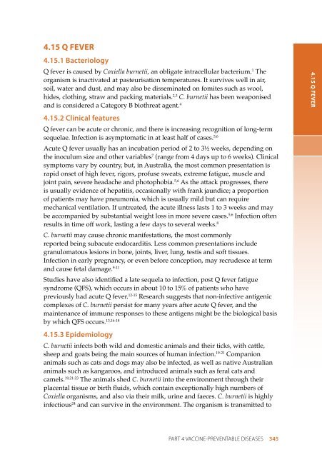
- Page 343 and 344: Children aged >5 years to 15 years)
- Page 345 and 346: Non-Indigenous adults A single dose
- Page 347 and 348: Adults who have a condition listed
- Page 349 and 350: 4.13.11 Adverse events 10-valent pn
- Page 351 and 352: 5 years with a condition(s) associa
- Page 353 and 354: virtually eradicated in India, but
- Page 355 and 356: formaldehyde, glutaraldehyde, polys
- Page 357: 4.14.8 Pregnancy and breastfeeding
- Page 361 and 362: 4.15.4 Vaccine • Q-Vax - CSL Limi
- Page 363 and 364: individuals, which can be accessed
- Page 365 and 366: Refer to 3.3 Groups with special va
- Page 367 and 368: 4.16 RABIES AND OTHER LYSSAVIRUSES
- Page 369 and 370: 4.16.4 Rabies vaccines • Mérieux
- Page 371 and 372: of an even more accelerated schedul
- Page 373 and 374: Pre-exposure prophylaxis for rabies
- Page 375 and 376: The relevant state/territory health
- Page 377 and 378: Although data are limited on the ef
- Page 379 and 380: Table 4.16.2: Post-exposure prophyl
- Page 381 and 382: Figure 4.16.2: Post-exposure prophy
- Page 383 and 384: Figure 4.16.3: Booster algorithm fo
- Page 385 and 386: allergic reaction occurs following
- Page 387 and 388: age group11,12 and affecting 3.8% o
- Page 389 and 390: of age, the risk of IS was increase
- Page 391 and 392: Interchangeability of rotavirus vac
- Page 393 and 394: gestational age; median 34 weeks) w
- Page 395 and 396: Infants living in households with p
- Page 397 and 398: 4.17.12 Variations from product inf
- Page 399 and 400: 2003, rubella notifications in Aust
- Page 401 and 402: zoster virus [Oka strain]). Lyophil
- Page 403 and 404: children
- Page 405 and 406: A number of commercial assays for t
- Page 407 and 408: Germany indicates that no case of v
- Page 409 and 410:
case. Seronegative women of child-b
- Page 411 and 412:
4.19 TETANUS 4.19.1 Bacteriology Te
- Page 413 and 414:
Formulations for children aged
- Page 415 and 416:
4.19.5 Transport, storage and handl
- Page 417 and 418:
foreign bodies (especially wood spl
- Page 419 and 420:
studies indicate that the adverse r
- Page 421 and 422:
The product information for Adacel
- Page 423 and 424:
of MDR-TB cases identified has incr
- Page 425 and 426:
BCG vaccination procedures BCG vacc
- Page 427 and 428:
Occupational groups There is some e
- Page 429 and 430:
4.20.13 Variations from product inf
- Page 431 and 432:
In developed countries, typhoid fev
- Page 433 and 434:
A 4th capsule taken on day 7 has be
- Page 435 and 436:
4.21.8 Pregnancy and breastfeeding
- Page 437 and 438:
4.22 VARICELLA 4.22.1 Virology Vari
- Page 439 and 440:
as a case of wild-type varicella oc
- Page 441 and 442:
Reconstituted Varivax Refrigerated
- Page 443 and 444:
adequate protection from varicella.
- Page 445 and 446:
eceived varicella vaccine while bre
- Page 447 and 448:
immunoglobulin and other blood prod
- Page 449 and 450:
eported in a 9-year follow-up of 70
- Page 451 and 452:
high-dose intravenous NHIG are like
- Page 453 and 454:
4.23 YELLOW FEVER 4.23.1 Virology Y
- Page 455 and 456:
Co-administration with other vaccin
- Page 457 and 458:
4.23.8 Pregnancy and breastfeeding
- Page 459 and 460:
Vaccine-associated neurotropic adve
- Page 461 and 462:
1000 cases per 100 000 population i
- Page 463 and 464:
administration of Zostavax with 23-
- Page 465 and 466:
diagnosis. In addition, the risk of
- Page 467 and 468:
Laboratory testing to check for an
- Page 469 and 470:
4.24.12 Variations from product inf
- Page 471 and 472:
the immunoglobulin preparations con
- Page 473 and 474:
Prevention of measles Measles vacci
- Page 475 and 476:
who are being treated with immunosu
- Page 477 and 478:
limb with a separate syringe, and a
- Page 479 and 480:
APPENDIX 1: CONTACT DETAILS FOR AUS
- Page 481 and 482:
APPENDIX 2: LITERATURE SEARCH STRAT
- Page 483 and 484:
APPENDIX 3: COMPONENTS OF VACCINES
- Page 485 and 486:
Vaccine component* Vaccine brand
- Page 487 and 488:
APPENDIX 4: COMMONLY ASKED QUESTION
- Page 489 and 490:
When should preterm infants be vacc
- Page 491 and 492:
If a parent decides not to have a c
- Page 493 and 494:
Should vaccines be given to persons
- Page 495 and 496:
eason not to vaccinate. Asthma, ecz
- Page 497 and 498:
Does MMR vaccine cause inflammatory
- Page 499 and 500:
(either alone or in combination) fo
- Page 501 and 502:
A4.5 Questions about the need for i
- Page 503 and 504:
APPENDIX 5: GLOSSARY OF TECHNICAL T
- Page 505 and 506:
Enzootic enzootic infections are pr
- Page 507 and 508:
Rotavirus a virus that is a common
- Page 509 and 510:
APPENDIX 6: COMMONLY USED ABBREVIAT
- Page 511 and 512:
OPV oral poliomyelitis vaccine PCEC
- Page 513 and 514:
Year Vaccine 2003 Varicella 2003 Me
- Page 515 and 516:
identifying the injection site, 79-
- Page 517 and 518:
Australian Capital Territory advers
- Page 519 and 520:
and travellers, 119 vaccines, 177-1
- Page 521 and 522:
abies and other lyssaviruses (inclu
- Page 523 and 524:
HPV Vaccination Program, 234 human
- Page 525 and 526:
interferon-gamma release assays (IG
- Page 527 and 528:
mercury, in vaccines. see thiomersa
- Page 529 and 530:
Northern Territory ACIR reporting,
- Page 531 and 532:
and Haemophilus influenzae type b (
- Page 533 and 534:
espiratory syncytial virus monoclon
- Page 535 and 536:
Therapeutic Goods Administration (T
- Page 537 and 538:
varicella-zoster immunoglobulin, 45
- Page 539 and 540:
INDEX 525 INDEX