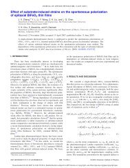
Rep. Prog. Phys. 73 (2010) 056502 The m<strong>in</strong>imum writable doma<strong>in</strong> size is not necessarily related to the <strong>in</strong>formation limit <strong>in</strong> PFM and can be either larger or smaller. This follows from the fact that while the signal generation volume <strong>in</strong> PFM is <strong>in</strong>dependent of the tip bias, the written doma<strong>in</strong> size, and <strong>in</strong> particular, the critical size of the nucleated doma<strong>in</strong>, has a strong bias dependence, i.e. m<strong>in</strong>imum writable doma<strong>in</strong> size can be smaller than the PFM resolution. This suggests that <strong>in</strong> some cases the resolution is a limit<strong>in</strong>g factor preclud<strong>in</strong>g experimental observation of smaller doma<strong>in</strong>s created by PFM. Clearly, this conclusion is non-universal and strongly depends on the material, e.g. <strong>in</strong> polycrystall<strong>in</strong>e films, the gra<strong>in</strong>-by-gra<strong>in</strong> switch<strong>in</strong>g will result <strong>in</strong> m<strong>in</strong>imal writable bit sizes be<strong>in</strong>g larger than the resolution. The resolution effect will clearly affect the analysis of the parameters such as doma<strong>in</strong> size distributions <strong>in</strong> the disordered <strong>materials</strong> or geometry of the fractal and self-aff<strong>in</strong>e doma<strong>in</strong> walls. For example, the structure factor will be S(q) = S ′ (q)R(q), where S ′ (q) is the <strong>in</strong>tr<strong>in</strong>sic structure factor of the <strong>in</strong>terface and R(q) is the transfer function def<strong>in</strong><strong>in</strong>g microscope resolution. Practically, R(q) = 1 for q ≪ q c and R(q) = q −n for q ≫ q c , where power law n is determ<strong>in</strong>ed by the image formation mechanism. For ultrath<strong>in</strong> <strong>in</strong>terfaces such as <strong>ferroelectric</strong> walls, q c ∼ 1/w c . Correspond<strong>in</strong>gly, the fractal properties h(x) for length scales below w c are likely dom<strong>in</strong>ated by the scanner noise along the slow scan axis (which can be established from the topographic imag<strong>in</strong>g of appropriate calibration standard, e.g. step edge of a cleaved graphite surface), and by the tails of the transfer function for the fast scan axis, rather than the <strong>in</strong>tr<strong>in</strong>sic wall geometry [219]. 3. <strong>Local</strong> <strong>polarization</strong> switch<strong>in</strong>g <strong>in</strong> <strong>ferroelectric</strong> <strong>materials</strong> by PFM 3.1. Polarization <strong>dynamics</strong> at the nanoscale The key characteristic of <strong>ferroelectric</strong> <strong>materials</strong> is that the direction of spontaneous <strong>polarization</strong> can be reversed by the application of sufficient electric field. Not surpris<strong>in</strong>gly, SPM of <strong>ferroelectric</strong>s has attracted considerable attention due to its potential to manipulate <strong>ferroelectric</strong> <strong>materials</strong> at the nanoscale by creat<strong>in</strong>g <strong>ferroelectric</strong> doma<strong>in</strong>s, study<strong>in</strong>g their <strong>dynamics</strong> dur<strong>in</strong>g growth and relaxation and mapp<strong>in</strong>g their <strong>in</strong>teraction with structural and morphological defects [107, 108, 110]. In this section, we summarize the exist<strong>in</strong>g results on the k<strong>in</strong>etics of doma<strong>in</strong> formation and relaxation, as well as theoretical models for the description of the doma<strong>in</strong> growth process <strong>in</strong> the rigid <strong>ferroelectric</strong> approximation. 3.2. Experimental aspects of tip-<strong>in</strong>duced <strong>polarization</strong> switch<strong>in</strong>g 3.2.1. Doma<strong>in</strong> growth k<strong>in</strong>etics. PFM allows a straightforward approach to study the k<strong>in</strong>etics of doma<strong>in</strong> formation and relaxation by comb<strong>in</strong><strong>in</strong>g the writ<strong>in</strong>g step of apply<strong>in</strong>g a bias pulse of preselected duration and magnitude, and the read<strong>in</strong>g stage at which the size of the result<strong>in</strong>g doma<strong>in</strong> is imaged. These studies have received a significant impetus <strong>in</strong> the context of <strong>ferroelectric</strong> data storage [25], and have been S V Kal<strong>in</strong><strong>in</strong> et al stimulated by the availability of high-quality epitaxial th<strong>in</strong> films that have low (below 10 V) switch<strong>in</strong>g voltages. A broad range of studies of doma<strong>in</strong> wall growth on sol– gel [220–223] <strong>ferroelectric</strong> films have been reported. The implementation of high-voltage PFM [224] has allowed studies of doma<strong>in</strong> <strong>dynamics</strong> <strong>in</strong> s<strong>in</strong>gle crystals as well, and particularly has enabled the k<strong>in</strong>etic studies as a function of pulse parameters [225–232]. As an example, shown <strong>in</strong> figure 22 is the morphology of <strong>ferroelectric</strong> doma<strong>in</strong>s <strong>in</strong> LNO s<strong>in</strong>gle crystal. While for low bias pulses the doma<strong>in</strong>s are nearly round, large doma<strong>in</strong>s adopt well-def<strong>in</strong>ed crystallographic shapes, mirror<strong>in</strong>g surface tension driven round<strong>in</strong>g of nanoparticles. The radii of doma<strong>in</strong>s fabricated <strong>in</strong> lithium niobate s<strong>in</strong>gle crystals were found to scale l<strong>in</strong>early with applied field and approximately logarithmically with time [226]. Similar scal<strong>in</strong>g was found for other <strong>materials</strong>, suggest<strong>in</strong>g the universality of this rate law. The time dependence of doma<strong>in</strong>-wall velocity was studied by several groups [128, 233, 234] <strong>in</strong> an attempt to relate doma<strong>in</strong> growth k<strong>in</strong>etics to the dom<strong>in</strong>ant wall p<strong>in</strong>n<strong>in</strong>g mechanisms. The first l<strong>in</strong>k between wall velocity and disorder was established <strong>in</strong> the sem<strong>in</strong>al papers by Tybell et al [64] and Paruch et al [234] and s<strong>in</strong>ce then was actively studied by several groups [233, 235]. Doma<strong>in</strong> growth <strong>in</strong> epitaxial films has also been compared with macroscopic measurements on capacitor structures [380]. Significant efforts have been directed at fabricat<strong>in</strong>g ultrasmall doma<strong>in</strong>s and the determ<strong>in</strong>ation of m<strong>in</strong>imal stable doma<strong>in</strong> size and associated lifetimes. Recently, 8 nm doma<strong>in</strong> arrays have been fabricated and detected us<strong>in</strong>g scann<strong>in</strong>g nonl<strong>in</strong>ear dielectric microscopy [237, 238] (figure 23(a)). The Cho group has also demonstrated the formation of 7 nm arrays and 2.8 nm doma<strong>in</strong>s (see figure 23(b)) and has written images us<strong>in</strong>g the technique (figure 23(c)) [236]. This impressive result corresponds to a density of 160 Tb <strong>in</strong>ch −2 (10 Tb <strong>in</strong>ch −2 for 8 nm array), approach<strong>in</strong>g molecular storage level [239]. One of the key uncerta<strong>in</strong>ties for the characterization of doma<strong>in</strong> growth k<strong>in</strong>etics is the lack of quantitative <strong>in</strong>formation on the electrostatic field structure produced by the probe. In most studies to date, the field is approximated as a s<strong>in</strong>gle po<strong>in</strong>t charge, a model well validated for tip apex at large separations from the contact area. On larger length scales, the conical part of the tip provides a slow-decay<strong>in</strong>g field component, which is, however, attenuated by the dielectric gap effect. Furthermore, the po<strong>in</strong>t charge model is clearly <strong>in</strong>applicable for small tip–surface separations of the order of the tip size (which <strong>in</strong> turn is related to the doma<strong>in</strong> wall width, as discussed <strong>in</strong> section 2.3, on which the field is uniform). The second factor affect<strong>in</strong>g doma<strong>in</strong> growth k<strong>in</strong>etics is the effect of mobile surface charges, screen<strong>in</strong>g and liquid bridge formation, as discussed <strong>in</strong> section 3.3. 3.2.2. Doma<strong>in</strong> relaxation and retention. The retention behavior of <strong>ferroelectric</strong> doma<strong>in</strong>s follow<strong>in</strong>g local <strong>polarization</strong> reversal presents obvious <strong>in</strong>terest for data storage and <strong>ferroelectric</strong> lithography applications. Retention behavior <strong>in</strong> epitaxial and polycrystall<strong>in</strong>e PZT [240], the thermal stability [228] and the retention behavior [229] of fabricated doma<strong>in</strong>s 23
Rep. Prog. Phys. 73 (2010) 056502 S V Kal<strong>in</strong><strong>in</strong> et al Figure 22. (a) PFM phase image of doma<strong>in</strong>s written <strong>in</strong> a lithium niobate crystal by vary<strong>in</strong>g the voltage pulse amplitude. Plots of doma<strong>in</strong> radius versus (b) pulse amplitude and (c) pulse duration <strong>in</strong> a lithium niobate crystal. (e) Doma<strong>in</strong> size as a function of pulse duration for a 370 Å thick PZT film. (d) Doma<strong>in</strong> wall speed as a function of the <strong>in</strong>verse applied electric field for three PZT films of different thicknesses. Panels (a)–(c) reproduced with permission from [226], copyright 2005, American Institute of Physics, and panels (d),(e) reproduced with permission from [64], copyright 2002, American Physical Society. Figure 23. (a) 8 nm array of doma<strong>in</strong>s, (b) 2.8 nm doma<strong>in</strong> and (c) nanoscale doma<strong>in</strong>s written to construct an image. (a) Reproduced from [52], copyright 2006 IOP Publish<strong>in</strong>g and (b), (c) from [236], copyright 2007, Institute of Pure and Applied Physics. 24