Local polarization dynamics in ferroelectric materials
Local polarization dynamics in ferroelectric materials
Local polarization dynamics in ferroelectric materials

You also want an ePaper? Increase the reach of your titles
YUMPU automatically turns print PDFs into web optimized ePapers that Google loves.
Rep. Prog. Phys. 73 (2010) 056502<br />
S V Kal<strong>in</strong><strong>in</strong> et al<br />
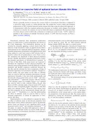
Figure 59. (a) Doma<strong>in</strong> structure of epitaxial PbZr 0.2 Ti 0.8 O 3 th<strong>in</strong> film with a pre-exist<strong>in</strong>g a 2 -positive doma<strong>in</strong> (<strong>polarization</strong> along [0 1 0])<br />
with<strong>in</strong> a matrix of c-negative doma<strong>in</strong> (<strong>polarization</strong> along [0 0 −1] under short-circuit boundary condition. The dots show the locations of the<br />
PFM tip along the profile A–M–N–B; (b) spatial distribution of nucleation potential at selected locations; (c) distribution of electrostatic<br />
energy density (MJ m −3 ) without applied potential [388]. Copyright 2008, American Institute of Physics.<br />
Figure 60. (a) Doma<strong>in</strong> structure of an (0 0 1)-oriented epitaxial PbZr 0.2 Ti 0.8 O 3 th<strong>in</strong> film; (b) spatial distribution of the nucleation potential<br />
along the profile P–Q–R–S–T–U–V [388]. Copyright 2008, American Institute of Physics.<br />
represents the nucleation potential correspond<strong>in</strong>g to a dot <strong>in</strong><br />
figure 59(a). It is shown that the potential required to nucleate<br />
a 180 ◦ doma<strong>in</strong> is lower near the ferroelastic tw<strong>in</strong> defects<br />
(∼1.6 V) as compared with ∼2.6 V away from the tw<strong>in</strong> defect<br />
with<strong>in</strong> the matrix. It is also found that the two parallel tw<strong>in</strong><br />
walls are not equivalent. The potential required to nucleate a<br />
180 ◦ doma<strong>in</strong> is lower near the left tw<strong>in</strong> boundary compared<br />
with the right one.<br />
The orig<strong>in</strong>s of the asymmetric variation of the nucleation<br />
voltage near the two tw<strong>in</strong> walls can be easily understood by<br />
analyz<strong>in</strong>g the electrostatic energy density on the surface of the<br />
th<strong>in</strong> film without any applied electric potential (figure 59(c)).<br />
The observation that the locations of the lowest nucleation<br />
voltage (figure 59(b)) do not co<strong>in</strong>cide with the locations of<br />
the tw<strong>in</strong> walls (po<strong>in</strong>t M or N <strong>in</strong> figure 59(c)) <strong>in</strong> the <strong>in</strong>itial<br />
doma<strong>in</strong> structure <strong>in</strong> figure 59(a) can be attributed to the<br />
<strong>in</strong>-plane electric field of PFM which slightly displaces the wall<br />
positions. F<strong>in</strong>ally, the small a-doma<strong>in</strong> size (∼10 nm) was not<br />
resolved s<strong>in</strong>ce the tip parameter γ was 30 nm, and hence only<br />
a s<strong>in</strong>gle large asymmetric dip <strong>in</strong> the nucleation voltage near<br />
the a-doma<strong>in</strong> is observed.<br />
5.6. Nucleation potential distribution <strong>in</strong> a doma<strong>in</strong> structure<br />
As shown above that the nucleation potential is different<br />
near ferroelastic tw<strong>in</strong> walls and with<strong>in</strong> a homogeneous<br />
doma<strong>in</strong> matrix, it is expected that the nucleation potential is<br />
<strong>in</strong>homogeneous with<strong>in</strong> a doma<strong>in</strong> structure. As an example,<br />
the spatial distribution of nucleation voltage was probed <strong>in</strong><br />
a more realistic doma<strong>in</strong> structure of PZT epitaxial th<strong>in</strong> film<br />
(figure 60(a))us<strong>in</strong>g phase-field simulations [388]. The doma<strong>in</strong><br />
structure is generated under a short-circuit boundary condition<br />
start<strong>in</strong>g from an <strong>in</strong>itial paraelectric state with small random<br />
perturbations. Each of the colors represents a tetragonal<br />
variant. The doma<strong>in</strong> structure consists of a 1 and a 2 doma<strong>in</strong>s<br />
embedded <strong>in</strong> a c-doma<strong>in</strong> matrix. To understand the correlation<br />
between the spatial variation of nucleation voltage and with<br />
the locations of <strong>ferroelectric</strong> tw<strong>in</strong> walls and wall junctions, the<br />
PFM tip position was moved along the l<strong>in</strong>e P–Q–R–S–T–U–V<br />
<strong>in</strong> figure 60(a). The nucleation voltage as a function of position<br />
is recorded <strong>in</strong> figure 60(b). It is observed that the nucleation<br />
potential is correlated with the number of local tw<strong>in</strong> doma<strong>in</strong><br />
variants. For example, the nucleation voltage is highest with<strong>in</strong><br />
the c-doma<strong>in</strong> matrix (po<strong>in</strong>ts R and U) followed by a s<strong>in</strong>gle tw<strong>in</strong><br />
wall (po<strong>in</strong>t S), and then by the area where a 1 and a 2 <strong>in</strong>tersect<br />
(po<strong>in</strong>t Q). The lowest nucleation potential is observed near the<br />
triple junctions (po<strong>in</strong>t T) where three doma<strong>in</strong>s meet.<br />
Remarkably, these examples illustrate that the comb<strong>in</strong>ation<br />
of the phase-field model<strong>in</strong>g and piezoresponse force<br />
microscopy and spectroscopy studies effectively allow us to<br />
study <strong>polarization</strong> <strong>dynamics</strong> at the level of a s<strong>in</strong>gle mesoscopic<br />
59