Abstracts - Association for Chemoreception Sciences
Abstracts - Association for Chemoreception Sciences
Abstracts - Association for Chemoreception Sciences
Create successful ePaper yourself
Turn your PDF publications into a flip-book with our unique Google optimized e-Paper software.
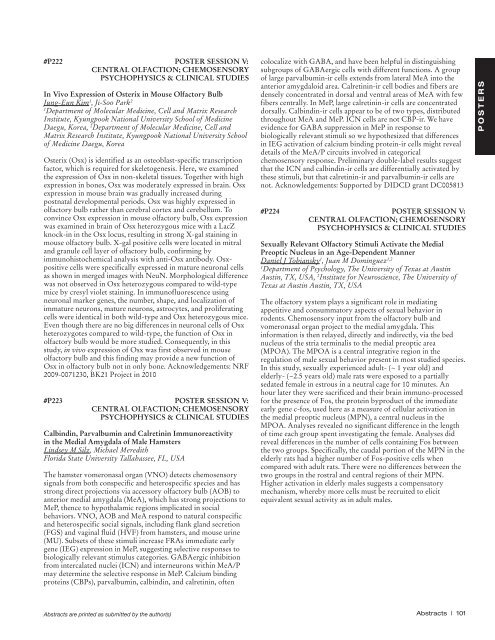
#P222 POSTER SESSION V:<br />
CENTRAL OLFACTION; CHEMOSENSORY<br />
PSYCHOPHYSICS & CLINICAL STUDIES<br />
In Vivo Expression of Osterix in Mouse Olfactory Bulb<br />
Jung-Eun Kim 1 , Ji-Soo Park 2<br />
1<br />
Department of Molecular Medicine, Cell and Matrix Research<br />
Institute, Kyungpook National University School of Medicine<br />
Daegu, Korea, 2 Department of Molecular Medicine, Cell and<br />
Matrix Research Institute, Kyungpook National University School<br />
of Medicine Daegu, Korea<br />
Osterix (Osx) is identified as an osteoblast-specific transcription<br />
factor, which is required <strong>for</strong> skeletogenesis. Here, we examined<br />
the expression of Osx in non-skeletal tissues. Together with high<br />
expression in bones, Osx was moderately expressed in brain. Osx<br />
expression in mouse brain was gradually increased during<br />
postnatal developmental periods. Osx was highly expressed in<br />
olfactory bulb rather than cerebral cortex and cerebellum. To<br />
convince Osx expression in mouse olfactory bulb, Osx expression<br />
was examined in brain of Osx heterozygous mice with a LacZ<br />
knock-in in the Osx locus, resulting in strong X-gal staining in<br />
mouse olfactory bulb. X-gal positive cells were located in mitral<br />
and granule cell layer of olfactory bulb, confirming by<br />
immunohistochemical analysis with anti-Osx antibody. Osxpositive<br />
cells were specifically expressed in mature neuronal cells<br />
as shown in merged images with NeuN. Morphological difference<br />
was not observed in Osx heterozygous compared to wild-type<br />
mice by cresyl violet staining. In immunofluorescence using<br />
neuronal marker genes, the number, shape, and localization of<br />
immature neurons, mature neurons, astrocytes, and proliferating<br />
cells were identical in both wild-type and Osx heterozygous mice.<br />
Even though there are no big differences in neuronal cells of Osx<br />
heterozygotes compared to wild-type, the function of Osx in<br />
olfactory bulb would be more studied. Consequently, in this<br />
study, in vivo expression of Osx was first observed in mouse<br />
olfactory bulb and this finding may provide a new function of<br />
Osx in olfactory bulb not in only bone. Acknowledgements: NRF<br />
2009-0071230, BK21 Project in 2010<br />
#P223 POSTER SESSION V:<br />
CENTRAL OLFACTION; CHEMOSENSORY<br />
PSYCHOPHYSICS & CLINICAL STUDIES<br />
Calbindin, Parvalbumin and Calretinin Immunoreactivity<br />
in the Medial Amygdala of Male Hamsters<br />
Lindsey M Silz, Michael Meredith<br />
Florida State University Tallahassee, FL, USA<br />
The hamster vomeronasal organ (VNO) detects chemosensory<br />
signals from both conspecific and heterospecific species and has<br />
strong direct projections via accessory olfactory bulb (AOB) to<br />
anterior medial amygdala (MeA), which has strong projections to<br />
MeP, thence to hypothalamic regions implicated in social<br />
behaviors. VNO, AOB and MeA respond to natural conspecific<br />
and heterospecific social signals, including flank gland secretion<br />
(FGS) and vaginal fluid (HVF) from hamsters, and mouse urine<br />
(MU). Subsets of these stimuli increase FRAs immediate early<br />
gene (IEG) expression in MeP, suggesting selective responses to<br />
biologically relevant stimulus categories. GABAergic inhibition<br />
from intercalated nuclei (ICN) and interneurons within MeA/P<br />
may determine the selective response in MeP. Calcium binding<br />
proteins (CBPs), parvalbumin, calbindin, and calretinin, often<br />
colocalize with GABA, and have been helpful in distinguishing<br />
subgroups of GABAergic cells with different functions. A group<br />
of large parvalbumin-ir cells extends from lateral MeA into the<br />
anterior amygdaloid area. Calretinin-ir cell bodies and fibers are<br />
densely concentrated in dorsal and ventral areas of MeA with few<br />
fibers centrally. In MeP, large calretinin-ir cells are concentrated<br />
dorsally. Calbindin-ir cells appear to be of two types, distributed<br />
throughout MeA and MeP. ICN cells are not CBP-ir. We have<br />
evidence <strong>for</strong> GABA suppression in MeP in response to<br />
biologically relevant stimuli so we hypothesized that differences<br />
in IEG activation of calcium binding protein-ir cells might reveal<br />
details of the MeA/P circuits involved in categorical<br />
chemosensory response. Preliminary double-label results suggest<br />
that the ICN and calbindin-ir cells are differentially activated by<br />
these stimuli, but that calretinin-ir and parvalbumin-ir cells are<br />
not. Acknowledgements: Supported by DIDCD grant DC005813<br />
#P224 POSTER SESSION V:<br />
CENTRAL OLFACTION; CHEMOSENSORY<br />
PSYCHOPHYSICS & CLINICAL STUDIES<br />
Sexually Relevant Olfactory Stimuli Activate the Medial<br />
Preoptic Nucleus in an Age-Dependent Manner<br />
Daniel J Tobiansky 1 , Juan M Dominguez 1,2<br />
1<br />
Department of Psychology, The University of Texas at Austin<br />
Austin, TX, USA, 2 Institute <strong>for</strong> Neuroscience, The University of<br />
Texas at Austin Austin, TX, USA<br />
The olfactory system plays a significant role in mediating<br />
appetitive and consummatory aspects of sexual behavior in<br />
rodents. Chemosensory input from the olfactory bulb and<br />
vomeronasal organ project to the medial amygdala. This<br />
in<strong>for</strong>mation is then relayed, directly and indirectly, via the bed<br />
nucleus of the stria terminalis to the medial preoptic area<br />
(MPOA). The MPOA is a central integrative region in the<br />
regulation of male sexual behavior present in most studied species.<br />
In this study, sexually experienced adult- (~ 1 year old) and<br />
elderly- (~2.5 years old) male rats were exposed to a partially<br />
sedated female in estrous in a neutral cage <strong>for</strong> 10 minutes. An<br />
hour later they were sacrificed and their brain immuno-processed<br />
<strong>for</strong> the presence of Fos, the protein byproduct of the immediate<br />
early gene c-fos, used here as a measure of cellular activation in<br />
the medial preoptic nucleus (MPN), a central nucleus in the<br />
MPOA. Analyses revealed no significant difference in the length<br />
of time each group spent investigating the female. Analyses did<br />
reveal differences in the number of cells containing Fos between<br />
the two groups. Specifically, the caudal portion of the MPN in the<br />
elderly rats had a higher number of Fos-positive cells when<br />
compared with adult rats. There were no differences between the<br />
two groups in the rostral and central regions of their MPN.<br />
Higher activation in elderly males suggests a compensatory<br />
mechanism, whereby more cells must be recruited to elicit<br />
equivalent sexual activity as in adult males.<br />
P O S T E R S<br />
<strong>Abstracts</strong> are printed as submitted by the author(s)<br />
<strong>Abstracts</strong> | 101


