MEDICINSKI GLASNIK
MEDICINSKI GLASNIK
MEDICINSKI GLASNIK

Create successful ePaper yourself
Turn your PDF publications into a flip-book with our unique Google optimized e-Paper software.
272<br />
Medicinski Glasnik, Volumen 6, Number 2, August 2009<br />
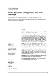
polyp creation in the right nasal cavity, situated<br />
in a lower nasal meatus and in left nasal cavity,<br />
obstructing the entire nasal cavity (Figure 1).<br />
Computed tomography (CT scan) of nasal<br />
cavity and paranasal sinuses in axial and coronary<br />
projection confirmed a shading of both sphenoid sinuses,<br />
partly of frontal ethmoids left, and both nasal<br />
cavities alongside unobstructed maxillary sinuses.<br />
With the powered instrument-assisted endoscopic<br />
approach under general endotracheal<br />
anesthesia, we identified a pearly polyp-creation<br />
centered between the middle nasal concha and<br />
a septum of the left nasal cavity with a petiole<br />
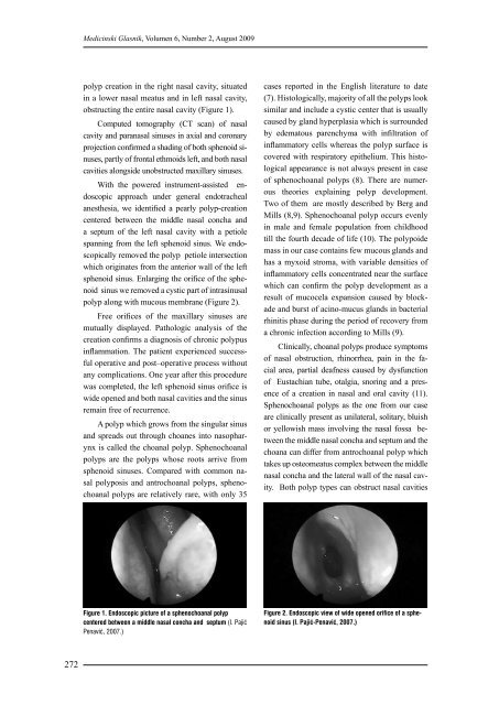
spanning from the left sphenoid sinus. We endoscopically<br />
removed the polyp petiole intersection<br />
which originates from the anterior wall of the left<br />
sphenoid sinus. Enlarging the orifice of the sphenoid<br />
sinus we removed a cystic part of intrasinusal<br />
polyp along with mucous membrane (Figure 2).<br />
Free orifices of the maxillary sinuses are<br />
mutually displayed. Pathologic analysis of the<br />
creation confirms a diagnosis of chronic polypus<br />
inflammation. The patient experienced successful<br />
operative and post–operative process without<br />
any complications. One year after this procedure<br />
was completed, the left sphenoid sinus orifice is<br />
wide opened and both nasal cavities and the sinus<br />
remain free of recurrence.<br />
A polyp which grows from the singular sinus<br />
and spreads out through choanes into nasopharynx<br />
is called the choanal polyp. Sphenochoanal<br />
polyps are the polyps whose roots arrive from<br />
sphenoid sinuses. Compared with common nasal<br />
polyposis and antrochoanal polyps, sphenochoanal<br />
polyps are relatively rare, with only 35<br />
Figure 1. Endoscopic picture of a sphenochoanal polyp<br />
centered between a middle nasal concha and septum (I. Pajić<br />
Penavić, 2007.)<br />
cases reported in the English literature to date<br />
(7). Histologically, majority of all the polyps look<br />
similar and include a cystic center that is usually<br />
caused by gland hyperplasia which is surrounded<br />
by edematous parenchyma with infiltration of<br />
inflammatory cells whereas the polyp surface is<br />
covered with respiratory epithelium. This histological<br />
appearance is not always present in case<br />
of sphenochoanal polyps (8). There are numerous<br />
theories explaining polyp development.<br />
Two of them are mostly described by Berg and<br />
Mills (8,9). Sphenochoanal polyp occurs evenly<br />
in male and female population from childhood<br />
till the fourth decade of life (10). The polypoide<br />
mass in our case contains few mucous glands and<br />
has a myxoid stroma, with variable densities of<br />
inflammatory cells concentrated near the surface<br />
which can confirm the polyp development as a<br />
result of mucocela expansion caused by blockade<br />
and burst of acino-mucus glands in bacterial<br />
rhinitis phase during the period of recovery from<br />
a chronic infection according to Mills (9).<br />
Clinically, choanal polyps produce symptoms<br />
of nasal obstruction, rhinorrhea, pain in the facial<br />
area, partial deafness caused by dysfunction<br />
of Eustachian tube, otalgia, snoring and a presence<br />
of a creation in nasal and oral cavity (11).<br />
Sphenochoanal polyps as the one from our case<br />
are clinically present as unilateral, solitary, bluish<br />
or yellowish mass involving the nasal fossa between<br />
the middle nasal concha and septum and the<br />
choana can differ from antrochoanal polyp which<br />
takes up osteomeatus complex between the middle<br />
nasal concha and the lateral wall of the nasal cavity.<br />
Both polyp types can obstruct nasal cavities<br />
Figure 2. Endoscopic view of wide opened orifice of a sphenoid<br />
sinus (I. Pajić-Penavić, 2007.)