MEDICINSKI GLASNIK
MEDICINSKI GLASNIK
MEDICINSKI GLASNIK

Create successful ePaper yourself
Turn your PDF publications into a flip-book with our unique Google optimized e-Paper software.
illary sinus, polypoid mucosa of ethmoid sinuses<br />
and orbital soft tissue swelling without focal abscess<br />
were found during a functional endoscopic<br />
surgery. All of necrotic tissue was removed and<br />
first histological examination showed chronic<br />
inflammation of paranasal sinusal mucosa. Bacterial<br />
and fungal cultures were negative. Four<br />
months later the patient developed fever, swelling,<br />
surface crusting, and widespread necrosis<br />
of the right periorbital and nasal area (Figure 2).<br />
Multiple biopsies of the paranasal sinuses were<br />
performed and diagnosed as nonspecific granulomatous<br />
inflammation.<br />
Finally, diagnosis of NK/T cell (CD 56+)<br />
lymphoma was made by histological and imunohistochemical<br />
reexamination of the paraffineembeded<br />
tissue obtained from the first biopsy of<br />
the ethmoid sinus and orbit. There were necrotic<br />
changes of varying degrees and a polymorphous<br />
pattern of proliferation involving large atypical<br />
cells with an occasional multilobated nucleus and<br />
various numbers of lymphocytes, plasma cells<br />
and macrophages. Features of vascular invasion<br />
by neoplastic lymphocytes were apparent. Occasionally,<br />
angiocentric pattern of proliferation was<br />
observed. Large atypical cells were positive for<br />
the NK-cell marker CD 56 (Figure 3). Patient had<br />
IV-A stage lymphoma and was EBV positive.<br />
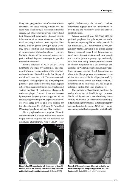
Neck lymph nodes were negative. Thoracic<br />
and abdominal CT scans as well as bone marrow<br />
biopsy were all negative. He was scheduled for<br />
continuous chemotherapy with 8 CHOP 14-day<br />
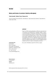
Figure 1. Axial CT scan showing soft tissue mass in the right<br />
ethmoid, frontal, and maxillary sinus eroding bony structures<br />
and infiltrating right medial rectus muscle (D. \anić, 2007.)<br />
Case report<br />
cycles. Unfortunately, the patient’s condition<br />
deteriorated rapidly after the development of<br />
liver failure and respiratory failure and after 18<br />
months he died.<br />
Primary paranasal sinus NK/T-cell (CD 56<br />
positive) lymphoma is a polymorphic extranodal<br />
lymphoma, expressing NK or rarely cytotoxic Tcell<br />
phenotype (3). It is an uncommon disease, and<br />
generally highly aggressive in its clinical course.<br />
Primary paranasal sinus T-cell lymphomas are<br />
much more frequent in Asian and Latin American<br />
countries, present at younger age, and usually<br />
arise from nasal cavity than the paranasal sinuses.<br />
In contrast, lymphomas of B-cell phenotype predominate<br />
in Western population and usually arise<br />
from paranasal sinuses. T-cell lymphomas are<br />
characterized by progressive ulceration and necrosis<br />
that are not typical for B-cell lymphomas (3, 5).<br />
Numerous studies showed that patients with NK/T<br />
lymphomas of the sinusonasal area had a high incidence<br />
of Epstein-Barr virus infections (6).<br />
The majority of lymphomas involving the<br />
ocular adnexa are of B-cell lineage. However,<br />
NK/T- cell lymphoma is associated only infrequently<br />
with orbital or adnexal involvement (7).<br />
Life style and environmental factors significantly<br />
increased risk for developing NK/T-cell lymphoma<br />
among individuals exposed to pesticides (8).<br />
Figure 2. Widespread necrosis of the right periorbital and<br />
nasal area in a patient with primary paranasal NK/T-cell<br />
lymphoma (D. \anić, 2007., with patient’s permission)<br />
275