- Page 1 and 2:
Propagation of Romulea species Pier
- Page 3 and 4:
Contents 2.7 GERMINATION PHYSIOLOGY
- Page 5 and 6:
Contents 5.3.1 Explants from seedli
- Page 7 and 8:
Abstract The suitability of various
- Page 9 and 10:
Declarations I Pierre André Swart,
- Page 11 and 12:
Declarations DETAILS OF CONTRIBUTIO
- Page 13 and 14:
Publications from this thesis ASCOU
- Page 15 and 16:
List of Figures Figure 2.13: Suther
- Page 17 and 18:
was significantly different from th
- Page 19 and 20:
List of Tables Table 2.1: Names of
- Page 21 and 22:
List of Abbreviations 2,4-D 2,4-Dic
- Page 23 and 24:
Die vlakhaas spits sy oor hy het di
- Page 25 and 26:
As die mens dan ook so sy dankbaarh
- Page 27 and 28:
Introduction where a great number o
- Page 29 and 30:
Introduction The subgenera Romulea
- Page 31 and 32:
2 Literature review Leef van daad e
- Page 33 and 34:
Literature review Figure 2.2: Life
- Page 35 and 36:
Literature review Figure 2.4: Life
- Page 37 and 38:
Literature review The plants are sh
- Page 39 and 40:
Literature review flowers (DE VOS,
- Page 41 and 42:
Literature review often found on cl
- Page 43 and 44:
Literature review flowers of R. lei
- Page 45 and 46:
Literature review flattened upper s
- Page 47 and 48:
2.2.16 Romulea tabularis Literature
- Page 49 and 50:
Literature review extinct, R. papyr
- Page 51 and 52:
Literature review R. leipoldtii occ
- Page 53 and 54:
Literature review Figure 2.8: Calvi
- Page 55 and 56:
Literature review Figure 2.14: Fras
- Page 57 and 58:
Figure 2.20: Nieuwoudtville weather
- Page 59 and 60:
Literature review Figure 2.26: Grah
- Page 61 and 62:
Literature review Figure 2.29: Diag
- Page 63 and 64:
Literature review Figure 2.30: A te
- Page 65 and 66:
Literature review Macronutrients ca
- Page 67 and 68:
Literature review The soils of the
- Page 69 and 70:
Literature review The embryo is the
- Page 71 and 72:
Literature review Phase 2 is seen a
- Page 73 and 74:
Literature review endogenous gibber
- Page 75 and 76:
Literature review tolerable level (
- Page 77 and 78:
Literature review these channels pe
- Page 79 and 80: Literature review 2.8.7 Seed dorman
- Page 81 and 82: Literature review the embryo (COPEL
- Page 83 and 84: Literature review germinated under
- Page 85 and 86: Literature review (COPELAND, 1976).
- Page 87 and 88: 2.9 BRIEF REVIEW OF IN VITRO CULTUR
- Page 89 and 90: Literature review (DEBERGH, 1994).
- Page 91 and 92: Literature review PIERIK (1997) sta
- Page 93 and 94: Literature review The distinguishin
- Page 95 and 96: Literature review The essential fun
- Page 97 and 98: 2.9.3.4 Abscisic acid Literature re
- Page 99 and 100: Table 2.6: Example of a matrix to e
- Page 101 and 102: Literature review Young embryos req
- Page 103 and 104: Literature review and the addition
- Page 105 and 106: Literature review organ is then pro
- Page 107 and 108: Literature review environmental str
- Page 109 and 110: 2.11 IN VITRO FLOWERING Literature
- Page 111 and 112: Literature review higher regenerati
- Page 113 and 114: Subfamily Ixioideae (continued) Tri
- Page 115 and 116: Literature review Table 2.8: Corm i
- Page 117 and 118: 3 Investigation into the habitat of
- Page 119 and 120: Soil sampling and analysis The firs
- Page 121 and 122: Soil sampling and analysis Table 3.
- Page 123 and 124: 4 Germination physiology 4.1 INTROD
- Page 125 and 126: 4.2.2 Water content and imbibition
- Page 127 and 128: Germination physiology (-N), phosph
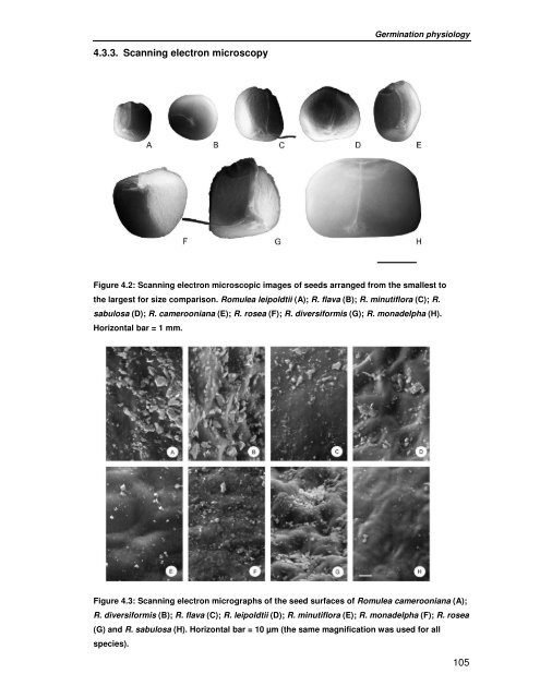
- Page 129: Germination physiology rosea seeds
- Page 133 and 134: Germination (%) 50 40 30 20 10 0 77
- Page 135 and 136: Germination (%) 100 80 60 40 20 0 c
- Page 137 and 138: Germination physiology The relative
- Page 139 and 140: Germination physiology seeds of R.
- Page 141 and 142: 5.2 MATERIALS AND METHODS In vitro
- Page 143 and 144: In vitro culture initiation and mul
- Page 145 and 146: 5.2.3 Explant comparison In vitro c
- Page 147 and 148: In vitro culture initiation and mul
- Page 149 and 150: In vitro culture initiation and mul
- Page 151 and 152: In vitro culture initiation and mul
- Page 153 and 154: In vitro culture initiation and mul
- Page 155 and 156: In vitro culture initiation and mul
- Page 157 and 158: In vitro culture initiation and mul
- Page 159 and 160: 5.4 DISCUSSION In vitro culture ini
- Page 161 and 162: In vitro culture initiation and mul
- Page 163 and 164: 6 In vitro corm formation and flowe
- Page 165 and 166: In vitro corm formation and floweri
- Page 167 and 168: 6.3 RESULTS 6.3.1 Corm formation In
- Page 169 and 170: In vitro corm formation and floweri
- Page 171 and 172: In vitro corm formation and floweri
- Page 173 and 174: In vitro corm formation and floweri
- Page 175 and 176: In vitro corm formation and floweri
- Page 177 and 178: Commercialization potential of Romu
- Page 179 and 180: Commercialization potential of Romu
- Page 181 and 182:
BASKIN, C. C., and BASKIN, J. M. (1
- Page 183 and 184:
Literature cited DOWLING, P. M., CL
- Page 185 and 186:
Literature cited HILTON-TAYLOR, C.
- Page 187 and 188:
Literature cited KUMAR, A., SOOD, A
- Page 189 and 190:
Literature cited PIERIK, R. L. M. (
- Page 191 and 192:
Literature cited STEINITZ, B., COHE