View/Open - ResearchSpace - University of KwaZulu-Natal
View/Open - ResearchSpace - University of KwaZulu-Natal
View/Open - ResearchSpace - University of KwaZulu-Natal

Create successful ePaper yourself
Turn your PDF publications into a flip-book with our unique Google optimized e-Paper software.
In vitro culture initiation and multiplication<br />
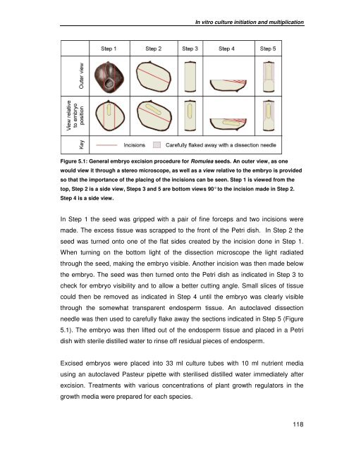
Figure 5.1: General embryo excision procedure for Romulea seeds. An outer view, as one<br />
would view it through a stereo microscope, as well as a view relative to the embryo is provided<br />
so that the importance <strong>of</strong> the placing <strong>of</strong> the incisions can be seen. Step 1 is viewed from the<br />
top, Step 2 is a side view, Steps 3 and 5 are bottom views 90° to the incision made in Step 2.<br />
Step 4 is a side view.<br />
In Step 1 the seed was gripped with a pair <strong>of</strong> fine forceps and two incisions were<br />
made. The excess tissue was scrapped to the front <strong>of</strong> the Petri dish. In Step 2 the<br />
seed was turned onto one <strong>of</strong> the flat sides created by the incision done in Step 1.<br />
When turning on the bottom light <strong>of</strong> the dissection microscope the light radiated<br />
through the seed, making the embryo visible. Another incision was then made below<br />
the embryo. The seed was then turned onto the Petri dish as indicated in Step 3 to<br />
check for embryo visibility and to allow a better cutting angle. Small slices <strong>of</strong> tissue<br />
could then be removed as indicated in Step 4 until the embryo was clearly visible<br />
through the somewhat transparent endosperm tissue. An autoclaved dissection<br />
needle was then used to carefully flake away the sections indicated in Step 5 (Figure<br />
5.1). The embryo was then lifted out <strong>of</strong> the endosperm tissue and placed in a Petri<br />
dish with sterile distilled water to rinse <strong>of</strong>f residual pieces <strong>of</strong> endosperm.<br />
Excised embryos were placed into 33 ml culture tubes with 10 ml nutrient media<br />
using an autoclaved Pasteur pipette with sterilised distilled water immediately after<br />
excision. Treatments with various concentrations <strong>of</strong> plant growth regulators in the<br />
growth media were prepared for each species.<br />
118