14th ICID - Poster Abstracts - International Society for Infectious ...
14th ICID - Poster Abstracts - International Society for Infectious ...
14th ICID - Poster Abstracts - International Society for Infectious ...

Create successful ePaper yourself
Turn your PDF publications into a flip-book with our unique Google optimized e-Paper software.
When citing these abstracts please use the following reference:<br />
Author(s) of abstract. Title of abstract [abstract]. Int J Infect Dis 2010;14S1: Abstract number.<br />
Please note that the official publication of the <strong>International</strong> Journal of <strong>Infectious</strong> Diseases 2010, Volume 14, Supplement 1<br />
is available electronically on http://www.sciencedirect.com<br />
Final Abstract Number: 34.004<br />
Session: Zoonoses and Infectoins in Animals<br />
Date: Wednesday, March 10, 2010<br />
Time: 12:30-13:30<br />
Room: <strong>Poster</strong> & Exhibition Area/Ground Level<br />
Type: <strong>Poster</strong> Presentation<br />
Arconobacterium pyogenes associated with pulmonary and submandibular lymph node<br />
abscessation in white tailed deer (Odocoileus virginiaus)<br />
R. Afifi 1 , J. M. Sleeman 2 , G.K. Saunders 3 , T. Kaur 3<br />
1 Afifi Department of Wildlife & Zoo Medicine, Faculty of Veterinary Medicine, Suez Canal<br />
University, Ismilia, Egypt, 2 Sleeman, USGS National Wildlife Health Center is located at 6006<br />
Schroeder Road, Madison, Wisconsin 53711, 3 Saunders,& Kaur, Department of Biomedical<br />
Sciences & Pathobiology, Virginia–Maryland Regional College of Veterinary Medicine, Virginia<br />
Polytechnic Institute and State University, Blacksburg, Virginia 24061, USA<br />
Background: Thin, lactating and uncoordinated female white-tailed deer was submitted <strong>for</strong><br />
necropsy as part of a surveillance program <strong>for</strong> chronic wasting disease (CWD). Laboratory tests<br />
<strong>for</strong> CWD and rabies were negative. Post-mortem examination revealed pulmonary and<br />
submandibular lymph node abscesses associated with Arcanobacterium pyogenes and<br />
Pasteurella. The overall presentation suggests that the infections may have been associated with<br />
chronic stress.<br />
Methods: Brain tissues were removed aseptically and transferred to Virginia’s Department of<br />
Game and Inland Fisheries <strong>for</strong> analysis <strong>for</strong> CWD and rabies virus, and Brain culture swabs were<br />
sent to the Virginia-Maryland Regional College of Veterinary Medicine <strong>for</strong> aerobic and anaerobic<br />
bacterial cultures, including Listeria monocytogenes culture. Lung, lymph node, brain, intestine<br />
and heart samples were preserved in 10% neutral buffered <strong>for</strong>malin <strong>for</strong> histopathologic<br />
examination. Lung and lymph node samples were taken aseptically <strong>for</strong> aerobic culture, including<br />
culture <strong>for</strong> Mycoplasma and Salmonella. Lung and lymph node were plated onto Blood Agar,<br />
MacConkey Agar and Columbia CAN Agar. Culture swabs of lung tissue were plated onto<br />
Chocolate agar, TSA and CAN agar. Chocolate Agar plates were incubated in 5% CO2 incubated<br />
at 37 ºC with no CO2 analysis.<br />
Results: Arcanobacterium pyogenes was isolated from lung and submandibular lymph node, and<br />
identified using bioMérieux API Coryne strips. Pasteurella spp. was isolated from the same lymph<br />
node, and identified using bioMérieux API 20 NE strip. Laboratory tests <strong>for</strong> rabies, CWD, Listeria,<br />
Mycoplasma, Mycobacterium and Salmonella were all negative. Histopathologic examination was<br />
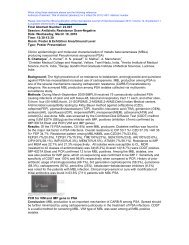
per<strong>for</strong>med on lung, brain, spleen, lymph node, intestine, heart and liver.The lung had multiple<br />
discrete nodules of coagulative necrosis containing neutrophils and macrophages (figure 1). A rim<br />
of neutrophilic inflammation surrounded the necrosis; peripheral to this was a layer of fibroplasia<br />
and fibrosis.<br />
Conclusion: In conclusion, even though it was not isolated from the lung, Pasteurella was the<br />
primary cause of infection in the lung and from there it spread to the lymph node. A. pyogenes<br />
was considered to be a secondary infection in the lungs where pneumonia was already present.<br />
The final diagnosis was pulmonary and lymph node abscesses due to A pyogenes and<br />
fibrinopurulent and necrotizing bronochopneumonia due to a mixed Pasteurella and A. pyogenes.