14th ICID - Poster Abstracts - International Society for Infectious ...
14th ICID - Poster Abstracts - International Society for Infectious ...
14th ICID - Poster Abstracts - International Society for Infectious ...

Create successful ePaper yourself
Turn your PDF publications into a flip-book with our unique Google optimized e-Paper software.
When citing these abstracts please use the following reference:<br />
Author(s) of abstract. Title of abstract [abstract]. Int J Infect Dis 2010;14S1: Abstract number.<br />
Please note that the official publication of the <strong>International</strong> Journal of <strong>Infectious</strong> Diseases 2010, Volume 14, Supplement 1<br />
is available electronically on http://www.sciencedirect.com<br />
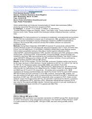
Final Abstract Number: 34.013<br />
Session: Zoonoses and Infectoins in Animals<br />
Date: Wednesday, March 10, 2010<br />
Time: 12:30-13:30<br />
Room: <strong>Poster</strong> & Exhibition Area/Ground Level<br />
Type: <strong>Poster</strong> Presentation<br />
The micro-adenomatous lesions associated with Lawsonia intracellularis in the pig intestine<br />
M. Sueyoshi, R. Uemura, H. Nagatomo<br />
University of Miyazaki, Miyazaki, Japan<br />
Background: Lawsonia intracellularis orally infects and causes marked hyperplasia of<br />
enterocytes in pigs. The infected intestinal wall makes thickening remarkable. This disease is<br />
called porcine proliferative enteropathy (PPE) or porcine intestinal adenomatosis(PIA). L.<br />
intracellularis was spreads all over the world and that the microbes were infected with pigs of a lot<br />
of farms have been reported. The characteristic pathological lesion of PIA is well known, but the<br />
pathogenesis mechanism is not clear. In this study, the localization of L. intracellularis and the<br />
mucosal lesions were investigated on the intestine with onset or healthy pigs.<br />
Methods: The histopathological examination of the intestines of the 25 poor-growth piglets and<br />
67 healthy pigs at the meat inspection station were examined by Hematoxylin-Eosin staining,<br />
Warthin-Starry(WS) staining and immunohistochemical(IHC) method used the anti-L.<br />
intracellularis antibody. In addition, it was examined detection of a specific gene of L.<br />
intracellularis by PCR method about an intestinal frozen-, a <strong>for</strong>malin- and a paraffin-specimen.<br />
Results: In necropsy, no thickened intestinal wall was found in 25 poor-growth piglets, however,<br />
in two of them, the typical PIA histological lesions were found in from the jejunum to the rectum.<br />
These cases were diagnosed as atypical PIA. In addition, in the other two of them, an islandshaped<br />
micro-PIA lesion was distributed in the intestinal mucosa. The comma-shaped small<br />
bacteria were observed by WS staining, and the antigens of L. intracellularis were detected with<br />
IHC methods in the enterocytes of the micro-adenomatous lesions. The antigen of L.<br />
intracellularis was also detected in the intact superficial enterocytes. In 67 healthy pigs, the<br />
thickened intestinal wall was not found macroscopically. However, the focal adenomatous lesions<br />
with clear boundaries were observed to three pigs in them. A specific gene of L. intracellularis<br />
was detected by PCR method in the intestinal frozen-, the <strong>for</strong>malin- and the paraffin-specimen.<br />
Conclusion: In this study, it was confirmed that there was atypical PIA, and the island-shaped or<br />
the focal adenomatous lesions were also distributed in a normal intestine macroscopically. The<br />
microlesions was suggested on the stage of an early infection or the subclinical infection with L.<br />
intracellularis.