- Page 1 and 2:
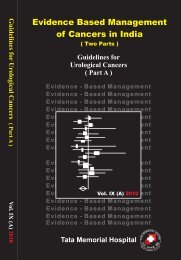
Guidelines forComplications ofCance
- Page 3:
Dedicated toAll our patients atThe
- Page 6 and 7:
Section IIHead & Neck 151Complicati
- Page 8 and 9:
PrefaceTreatment of cancer by vario
- Page 10:
Section — IGeneralContributorsDr.
- Page 13 and 14:
6. Complications after breast recon
- Page 15 and 16:
promises to reduce this risk of ser
- Page 17 and 18:
preceded by increased sensory hyper
- Page 19 and 20:
had significant lymphoedema. The fa
- Page 21 and 22:
2. Smoking as a risk factor for wou
- Page 23 and 24:
4. Preoperative core needle biopsy
- Page 25 and 26:
eduction mammoplasty, and axillary-
- Page 27 and 28:
postoperative wound infection follo
- Page 29 and 30:
ablative surgical treatment for car
- Page 31 and 32:
postoperative seroma formation usin
- Page 33 and 34:
dressing consisted of a circumferen
- Page 35 and 36:
mastectomies with axillary node cle
- Page 37 and 38:
mastectomy for breast cancer. Early
- Page 39 and 40:
monitored for the development of po
- Page 41 and 42:
modality, and tumor stage distribut
- Page 43 and 44:
26. Pain and other symptoms during
- Page 45 and 46:
28. Coping, catastrophizing and chr
- Page 47 and 48:
surgical technique at the time of m
- Page 49 and 50:
0.001), 0.315 cm +/- 1.27 at the an
- Page 51 and 52:
or heat and superficial incision is
- Page 53 and 54:
RecommendationsA. Preparation of th
- Page 55 and 56:
3. Mangram AJ, Horan TC, Pearson ML
- Page 57 and 58:
Mild hypothermia increases blood lo
- Page 59 and 60:
Perioperative Normothermia to Reduc
- Page 61 and 62:
Priorities: Restoration of circulat
- Page 63 and 64:
than 1.5 times control values but f
- Page 65 and 66:
outcomes that may result from the u
- Page 67 and 68:
Preoperative Preparation of the Pat
- Page 69 and 70:
patients, or other patients who may
- Page 71 and 72:
Obstruction can occur at the level
- Page 74 and 75:
c. Administer face mask preoxygenat
- Page 76 and 77:
5. The use of exhaled carbon dioxid
- Page 78 and 79:
disorder) may occur after an episod
- Page 80 and 81:
undergoing general anesthesia, eith
- Page 82 and 83:
anesthesia awareness occurred in ea
- Page 84 and 85:
The current guidelines are actually
- Page 86 and 87:
o S - Strong Recommendation -Commit
- Page 88 and 89:
oSource control ASAP after successf
- Page 90 and 91:
oooWean steroids once vasopressors
- Page 92 and 93:
Mechanical ventilation of sepsis-in
- Page 94 and 95:
o CVVH offers easier management inh
- Page 96 and 97:
critically ill patient that are con
- Page 98 and 99:
Rivers E, Nguyen B, Havstad S, et a
- Page 100 and 101:
in survival of 7.6%. By the second
- Page 102 and 103: parameters (gastric mucosal Pco2, s
- Page 104 and 105: syndrome. We therefore conducted a
- Page 106 and 107: Complications After Limb SalvageSur
- Page 108 and 109: local tumor and to preserve as much
- Page 110 and 111: femoral head to the center of the k
- Page 112 and 113: 4. Griffin AM, Parsons JA, Davis AM
- Page 114 and 115: 20. Roberts P, Chan D, Grimer RJ, e
- Page 116 and 117: An infection can have disastrous co
- Page 118 and 119: and facilitating reimplantation sur
- Page 120 and 121: features of a two-stage revision is
- Page 122 and 123: patients had an amputation. Two oth
- Page 124 and 125: its main role is confined to ruling
- Page 126 and 127: 2. Capanna R, Morris HG, Campanacci
- Page 128 and 129: 21. Wilson MG, Kelley K, Thornhill
- Page 130 and 131: 39. Love C, Marwin SE, Tomas MB, Kr
- Page 132 and 133: 5. A systematic review of 25 RCT’
- Page 134 and 135: (cefazolin or cefuroxime) or penici
- Page 136 and 137: should be based on the isolates fro
- Page 138 and 139: such as concomitant use of prophyla
- Page 140 and 141: Surgical techniqueAdherence to meti
- Page 142 and 143: 2. Another small prospective random
- Page 144 and 145: Antibiotic prophylaxis for dental a
- Page 146 and 147: Antimicrobial Prophylaxis in Orthop
- Page 148 and 149: 35. Tai CC, Nirvani AA, Holmes A, e
- Page 150 and 151: 53. Parker MJ, Roberts C. Closed su
- Page 154 and 155: sequences may be of value. They cau
- Page 156 and 157: an excellent alternative to amputat
- Page 158: 5. Hünerbein M, Hohenberger P, Str
- Page 162 and 163: Oral IncompetenceOral competence is
- Page 164 and 165: flap is insensate and has no motor
- Page 166 and 167: 16. R. Song, Y. Gao, Y. Song, Y. Yu
- Page 168 and 169: The incidence is usually lower afte
- Page 170 and 171: Late fibrotic phase: Attempted tiss
- Page 172 and 173: Ref: A Lyons, N Ghazali/ British J
- Page 174 and 175: 17. Store G, Boysen M. Mandibular o
- Page 176 and 177: amino acids, proteins and traces of
- Page 178 and 179: RTOG, EORTC and CTCAE, version 3 fo
- Page 180 and 181: difference in the disease related p
- Page 182 and 183: References1. Bourhis J, Rosine D. R
- Page 184 and 185: with head and neck cancer. N Engl J
- Page 186 and 187: MucositisMucositis is an inflammato
- Page 188 and 189: Type of cancer: high risk for mucos
- Page 190 and 191: Oral care protocol1. Brush all toot
- Page 192 and 193: 3. El-Sayed S, Nabid A, ShelleyW, e
- Page 194 and 195: 1) Recipient site problems- The maj
- Page 196 and 197: estoration of vascular flow to tiss
- Page 198 and 199: General Complications- Deep vein th
- Page 200 and 201: Nerve Injuries(Recurrent and Superi
- Page 202 and 203:
paramedian position, it will result
- Page 204 and 205:
along with its variations, and a me
- Page 206 and 207:
anatomy as related to surgery of th
- Page 208 and 209:
Risk factorsCentral compartment (Le
- Page 210 and 211:
Goal - To raise serum calcium level
- Page 212 and 213:
Pharyngocutaneous Fistula (PC Fistu
- Page 214 and 215:
Based on our institutional practice
- Page 216 and 217:
fistula: The Memorial University ex
- Page 218 and 219:
Risk factors (LOE-5)Patient factors
- Page 220 and 221:
oCircumferencial excision and inser
- Page 222 and 223:
y trauma or tumor, exposing the und
- Page 224 and 225:
Arytenoid cartilage is reported to
- Page 226 and 227:
Filntisis, G.A., et al. “Laryngea
- Page 228 and 229:
As high as 52% patients are detecte
- Page 230 and 231:
ManagementPostural exercisesDisorde
- Page 232 and 233:
References1) Objective assessment o
- Page 234 and 235:
Diagnosis Average time to hypothyro
- Page 236 and 237:
Management of Facial Nerve InjuryIn
- Page 238 and 239:
2. Interposition or cable graft (to
- Page 240 and 241:
Prevention of corneal exposure Tars
- Page 242 and 243:
flushing) and localized sweating (g
- Page 244 and 245:
Other options :1. Intracutaneous in
- Page 246 and 247:
Shoulder Dysfunction or PainfulShou
- Page 248 and 249:
EMG studies on scapulo-humeral and
- Page 250 and 251:
Chyle Fistula after Neck Dissection
- Page 252 and 253:
Rationale:LCTs, which constitute 70
- Page 254 and 255:
CSF Leak after Craniofacial Resecti
- Page 256 and 257:
to the muscle to recreate the orbit
- Page 258 and 259:
Conservative treatmentIt is logical
- Page 260 and 261:
Complications Associated with use o
- Page 262 and 263:
Following is the example of hematol
- Page 264 and 265:
ooooooo direct interaction with the
- Page 266 and 267:
ooooointravenously over 15 minutes
- Page 268 and 269:
in most of the patients. The common
- Page 270:
Section — IIIRadiotherapyContribu
- Page 273 and 274:
nodes are involved or suspected to
- Page 275 and 276:
function. In general, with conventi
- Page 277 and 278:
like ultrasonography, CT scan, endo
- Page 279 and 280:
catheter passed through the anus in
- Page 281 and 282:
Acute symptoms usually subside seve
- Page 283 and 284:
granulocytopenia, thrombocytopenia
- Page 285 and 286:
adiation therapy), IMRT (intensity-
- Page 287 and 288:
patients who have received radical
- Page 289 and 290:
ate expected in the standard popula
- Page 292 and 293:
adjusting for competing causes of d
- Page 294 and 295:
therapy (3D-CRT) and intensity modu
- Page 296 and 297:
attributed to use of cisplatin base
- Page 298 and 299:
Conclusion:Incidence of SMN is incr
- Page 300 and 301:
11. Travis LB, Fossa SD, Schonfeld
- Page 302 and 303:
adiation-related solid SMNs will in
- Page 304 and 305:
0.71, p
- Page 306 and 307:
5.65%) within 45 years after a CNS
- Page 308 and 309:
adiation damage though supporting v
- Page 310 and 311:
without calcification. Inflammation
- Page 312 and 313:
patients treated using the original
- Page 314 and 315:
incidence of valvular thickening at
- Page 316 and 317:
12. Hancock S, Hoppe R. Long-term c
- Page 318 and 319:
syndrome and bronchiolitis oblitera
- Page 320 and 321:
Darby SC, McGale P, Taylor CW, Peto
- Page 322 and 323:
undertaken, based on individual pat
- Page 324 and 325:
Pathophysiological Aspects:The func
- Page 326 and 327:
Symptoms: Symptoms related to pulmo
- Page 328 and 329:
Prevention of Pneumonitis and Lung
- Page 330 and 331:
genotyping analysis of single nucle
- Page 332 and 333:
Registry of Patients. We computed I
- Page 334 and 335:
eceived elective nodal irradiation
- Page 336 and 337:
Int J Radiat Oncol Biol Phys. 1995
- Page 338 and 339:
worsening during the treatment shou
- Page 340 and 341:
Differential Diagnosis: This sign i
- Page 342 and 343:
Dose Incidence Comments Author>30 G
- Page 344 and 345:
PET scan findings have been describ
- Page 346 and 347:
3. Pallis CA, Louis S, Morgan RL.:
- Page 348 and 349:
24. Abbatucci JS, Delozier T, Quint
- Page 350 and 351:
and summarized into specific recomm
- Page 352 and 353:
adequate. With the increasing use o
- Page 354 and 355:
Complications are mainly described
- Page 356 and 357:
IQ decline is associated with sever
- Page 358 and 359:
activity’ of a person, essentiall
- Page 360 and 361:
development after brain tumor thera
- Page 362 and 363:
Highest relative risk of second can
- Page 364 and 365:
Suggested Reading:A) Neuropsycholog
- Page 366 and 367:
aseline. Male sex, longer symptomat
- Page 368 and 369:
number of patients. Cognition was p
- Page 370 and 371:
16 years, the corresponding value w
- Page 372 and 373:
15 of 20 (75%) subjects, endocrine
- Page 374 and 375:
OBJECTIVE: To assess cerebrovascula
- Page 376 and 377:
hypometabolism. Of the 17 patients
- Page 378:
10-20 months, with an average of 18
- Page 382 and 383:
Febrile NeutropeniaNeutropenia is w
- Page 384 and 385:
Approach to initial antibiotic use
- Page 386 and 387:
Known colonization with Penicillin/
- Page 388 and 389:
DocumentedinfectionsSuggesteddurati
- Page 390 and 391:
Zygomycosis and other mold infectio
- Page 392 and 393:
Overall risk Examples Febrile neutr
- Page 394 and 395:
Broad spectrum DOSE Spectrum Commen
- Page 396 and 397:
Broad spectrum DOSE Spectrum Commen
- Page 398 and 399:
Broad spectrum DOSE Spectrum Commen
- Page 400 and 401:
Role of G-CSF-Primary Prophylaxis:R
- Page 402 and 403:
Chemotherapy Induced Nausea andVomi
- Page 404 and 405:
Level I Level II Level III Level IV
- Page 406 and 407:
Types & doses of AntiemeticsDrug &
- Page 408 and 409:
supported by a meta-analysis, which
- Page 410 and 411:
and follow-up after treatment for n
- Page 412 and 413:
day 1; aprepitant and dexamethasone
- Page 414 and 415:
2-3: APR 80 mg every day) or a cont
- Page 416 and 417:
Late Effects in Childhood CancerSur
- Page 418 and 419:
to increase their awareness about l
- Page 420 and 421:
Contd...Score2BStatement of consens
- Page 422 and 423:
6 Kurkure PA, Achrekar S, Uparkar U
- Page 424 and 425:
nonirradiated group did not differ
- Page 426 and 427:
Gaslini Children’s Research Hospi
- Page 428 and 429:
Our ACT model has 3 basic facets; p
- Page 430 and 431:
The relative risk of developing TLS
- Page 432 and 433:
the excretion of uric acid and phos
- Page 434 and 435:
hyperuricemia (> 7.5 mg/dL), treatm
- Page 436 and 437:
hydration, and the administration o
- Page 438 and 439:
which the different drugs are admin
- Page 440 and 441:
Table 2. Cairo-Bishop clinical Tumo
- Page 442 and 443:
Table 4 Patient Stratification by R
- Page 444 and 445:
Table 6 Management of Electrolyte A
- Page 446 and 447:
of TLS and improve the management o
- Page 448 and 449:
4. Efficacy and safety of rasburica
- Page 450 and 451:
Pulmonary Toxicity of Antineoplasti
- Page 452 and 453:
Though there are many chemotherapeu
- Page 454 and 455:
corticosteroids with variable respo
- Page 456 and 457:
Nephrotoxicity ofChemotherapeutic D
- Page 458 and 459:
associated with use of carbopltin c
- Page 460 and 461:
Table 2 Chemotherapeutics Requiring
- Page 462 and 463:
against ifosfamide nephrotoxicity.
- Page 464 and 465:
The incidence of mucositis involvin
- Page 466 and 467:
7. Obesity - Probably due to over d
- Page 468 and 469:
conditioning treatment and for 3 da
- Page 470 and 471:
References and Recommended Readings
- Page 472 and 473:
metabolism for activity, cyclophosp
- Page 474 and 475:
delays excretion and eventually res
- Page 476 and 477:
Combination Chemotherapy:Combinatio
- Page 478 and 479:
Vincristine: No dose reduction if b
- Page 480 and 481:
19. Slichenmyer WJ, Rowinsky EK, Gr
- Page 482 and 483:
AlopeciaCommon drugs causing alopec
- Page 484 and 485:
dependent, occurring from 8 days to
- Page 486 and 487:
Cancer Induced Anemia (CIA)Introduc
- Page 488 and 489:
system. There is an upregulation of
- Page 490 and 491:
time may represent increasing anemi
- Page 492 and 493:
(rHuEPO) whiles the other received
- Page 494 and 495:
Table 3. Improvements in Hemoglobin
- Page 496 and 497:
2. Groopman JE, Itri LM. Chemothera
- Page 498 and 499:
epoetin alfa versus oral iron or no
- Page 500 and 501:
NOTES


