- Page 1 and 2: EUROPEANCOMMISSIONEuropeanResearch
- Page 3 and 4: EUROPEAN COMMISSIONINSPIRING RESEAR
- Page 5 and 6: Marie Curie Actions: Inspiring Rese
- Page 7 and 8: ContentsIntroduction . . . . . . .
- Page 9: WORLDWIDE RESEARCH . . . . . . . .
- Page 12 and 13: within the best European research g
- Page 14 and 15: Cross-border cooperation is the cor
- Page 16 and 17: The Marie Curie programme seeks to
- Page 18 and 19: AQUACHEMWater is life. It is key to
- Page 20 and 21: ground for early-stage and experien
- Page 22 and 23: CARBIOCarbon nanotubes are the epit
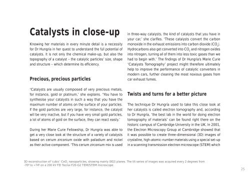
- Page 24 and 25: e chemically modifi ed to further e
- Page 28 and 29: included a specimen holder inserted
- Page 30 and 31: CRONUS-EUThe Earth is under attack:
- Page 32 and 33: led to an unprecedented ability to
- Page 34 and 35: HETEROLICSThe days of grainy graphi
- Page 36 and 37: Other researchers from Ural State T
- Page 38 and 39: HYDROGEN 1S-2SAccurate clocks are c
- Page 40 and 41: more accurate. Such a clock could b
- Page 42 and 43: RAPTOR ECOTOXFears over species los
- Page 44 and 45: in Sicily, as well as rock climbers
- Page 46 and 47: SMARTEverything about this material
- Page 48 and 49: Development scheme as a chance to m
- Page 50 and 51: SPATSTATHow can statistical analysi
- Page 52 and 53: Hristopulos. There are two categori
- Page 54 and 55: TRACKSResearch within the Tracks pr
- Page 56 and 57: intellectual property issues and th
- Page 58 and 59: Marie Curie Fellowships act as a sp
- Page 60 and 61: The seeds of a budding career are p
- Page 62 and 63: ARTEMISUnderstanding nature at its
- Page 64 and 65: network institutes have allowed the
- Page 66 and 67: BIOMEMA membrane is a layer of mate
- Page 68 and 69: Mighty Mini-MBABut Biomem also step
- Page 70 and 71: COMODEWhen consumers buy a new coff
- Page 72 and 73: with computer-aided design (CAD) to
- Page 74 and 75: COOPERATIVE BREEDINGCan our sibling
- Page 76 and 77:
mammals. Other analyses of cooperat
- Page 78 and 79:
CSIMHVALVESImagine being able to un
- Page 80 and 81:
could be used to describe how carti
- Page 82 and 83:
GLUESDr Markus Reichstein is a ‘r
- Page 84 and 85:
The carbon cycle begins with plants
- Page 86 and 87:
INTERDECHow an organism develops de
- Page 88 and 89:
The heart of the matterThe PhD Fell
- Page 90 and 91:
KAPROWThe theory of democratic peac
- Page 92 and 93:
necessary to stop it.’ If we are
- Page 94 and 95:
NASCENTAlthough nanotechnology deal
- Page 96 and 97:
The network focuses on the developm
- Page 98 and 99:
NEUROEMPATHYEver sense a chill runn
- Page 100 and 101:
Mapping empathySince the 1890s, it
- Page 102 and 103:
© stock.xchngOESTROGEN RECEPTORThe
- Page 104 and 105:
says Dr López-Béjar. Underlining
- Page 106 and 107:
PAPHOSIn 1870, Jules Verne wrote ab
- Page 108 and 109:
Minuscule communities under the sea
- Page 110 and 111:
PENELOPEPenelope, the wife of Ulyss
- Page 112 and 113:
Training and travelFor the Fellows,
- Page 114 and 115:
PUMPING UP THE HEARTMany researcher
- Page 116 and 117:
iochemical reactions take place in
- Page 118 and 119:
SEMEAIOutside the obvious green job
- Page 120 and 121:
suited to some companies than to ot
- Page 122 and 123:
Increasing our knowledge of the uni
- Page 124 and 125:
Everybody benefi ts from knowledge
- Page 126 and 127:
CABMarie Curie Fellows working unde
- Page 128 and 129:
The project achieved some major mil
- Page 130 and 131:
ENZYMATEnzymes are biomolecules tha
- Page 132 and 133:
eports. And the industry seems to s
- Page 134 and 135:
NUCSYSComplex organisms such as hum
- Page 136 and 137:
Integrating the ‘-omics’Nuclear
- Page 138 and 139:
TARGET-BREASTThe emotional turmoil
- Page 140 and 141:
diagnostics within an industrial en
- Page 142 and 143:
TEAMOHOLICAlthough it deals with an
- Page 144 and 145:
Dream teamAs its name suggests, Tea
- Page 146 and 147:
Women researchers have brought majo
- Page 148 and 149:
more likely to leave once they are
- Page 150 and 151:
AQUABASEToday, an increasingly comp
- Page 152 and 153:
‘With C14 isotopes, we can track
- Page 154 and 155:
DRUG-DISCOVERYDuring her two-year M
- Page 156 and 157:
chemically in various fashions. The
- Page 158 and 159:
EARA-ESTThe European Association fo
- Page 160 and 161:
‘Our programme is founded on the
- Page 162 and 163:
GLADNETAccurate data about the qual
- Page 164 and 165:
Working with industryIn addition to
- Page 166 and 167:
MIGRANT CHILDRENImmigration has bec
- Page 168 and 169:
Ireland possesses a category of mig
- Page 170 and 171:
NANBIOPTICOne of the most exciting
- Page 172 and 173:
Marie Curie support allowed me, amo
- Page 174 and 175:
NANOMATCHDeveloping new and more ef
- Page 176 and 177:
‘The ombudswoman is used quite fr
- Page 178 and 179:
Europe is increasingly retaining it
- Page 180 and 181:
well as the researcher’s scientif
- Page 182 and 183:
ADAPTUNPREDICTThe Marie Curie progr
- Page 184 and 185:
we understand the consequences bett
- Page 186 and 187:
© stock.xchngCSS-OMICSStress is a
- Page 188 and 189:
A key part of her research was to f
- Page 190 and 191:
ELSAQUINTANA-NCSCSFor Spanish pharm
- Page 192 and 193:
The cancer stem cell hypothesis pre
- Page 194 and 195:
EXNER_BORISOVMany modern devices an
- Page 196 and 197:
chips, for instance. ‘Of course,
- Page 198 and 199:
HIVOXFORDIn late 2006, five Bulgari
- Page 200 and 201:
that the accused medical personnel
- Page 202 and 203:
© stock.xchngMARINE MAGNETISMIsn
- Page 204 and 205:
aeolian dust. The second traces dus
- Page 206 and 207:
MODPAPThe Modpap project has delive
- Page 208 and 209:
According to Dr Macpherson, Dr Pill
- Page 210 and 211:
NANOSWITCHIt calls to mind a scene
- Page 212 and 213:
‘Throughout their journey, the na
- Page 214 and 215:
PIPKIGKOOne in every 700 babies bor
- Page 216 and 217:
focal adhesions of specifi c protei
- Page 218 and 219:
TARGETING PCD IN PDA Marie Curie gr
- Page 220 and 221:
the blood-brain barrier, the precur
- Page 222 and 223:
Science completely shapes the way w
- Page 224 and 225:
as labs and microscopes in order to
- Page 226 and 227:
BCRThe moon holds a strange fascina
- Page 228 and 229:
A new perspective on chronobiologyT
- Page 230 and 231:
© stock.xchngBIMORESolar energy is
- Page 232 and 233:
convert CO 2into a reduced carbon b
- Page 234 and 235:
CENS-CMAAs the Estonian coastline c
- Page 236 and 237:
The research visitors provided valu
- Page 238 and 239:
FLOODSVery few of us now question t
- Page 240 and 241:
The hunting ground for this preciou
- Page 242 and 243:
NANOMAG-LABThese days, the average
- Page 244 and 245:
What is more, Nanomag-Lab fostered
- Page 246 and 247:
NEPHILID SPIDERSSlovenian scientist
- Page 248 and 249:
One simple strategy involves the ma
- Page 250 and 251:
© stock.xchngWISEThe 32 countries
- Page 252 and 253:
and-tested methodology also include
- Page 254 and 255:
The quest for knowledge is a univer
- Page 256 and 257:
Global partnershipsBy opening up Eu
- Page 258 and 259:
ATTACCOnly one in a million is affe
- Page 260 and 261:
nutrients and ensures blood supply
- Page 262 and 263:
BIOSEBOut in the middle of nowhere,
- Page 264 and 265:
free discussion evenings, and staff
- Page 266 and 267:
CAUSE KIDNEY DAMAGEA potentially de
- Page 268 and 269:
might contribute to the effect. ‘
- Page 270 and 271:
CAVEOLINSEvery breath becomes a whe
- Page 272 and 273:
‘Our research revealed that the c
- Page 274 and 275:
EPISCONThe conservation and preserv
- Page 276 and 277:
able to follow whatever course my c
- Page 278 and 279:
© stock.xchngPARSEMUnder the Marie
- Page 280 and 281:
The research objectives of Parsem w
- Page 282 and 283:
NEURAL CIRCUITBlind mice can now se
- Page 284 and 285:
An international effortRecognising
- Page 286 and 287:
Being able to simultaneously build
- Page 288 and 289:
A number of initiatives can be impl
- Page 290 and 291:
EURYTHRONYoung researchers working
- Page 292 and 293:
Flexible work environmentThe resear
- Page 294 and 295:
INNATE IMMUNITYThe Innate Immunity
- Page 296 and 297:
more cytokines. Normally, this feed
- Page 298 and 299:
NLRS IN IMMUNITYMore than a million
- Page 300 and 301:
organisms and to shed light on the
- Page 302 and 303:
ZSN-PHD PROGRAMMEIt takes talent, t
- Page 304 and 305:
eview.’ The ZSN has also encourag
- Page 306 and 307:
The Marie Curie Actions are founded
- Page 308:
All this is designed to achieve one
- Page 311 and 312:
On soft groundWhen faced with groun
- Page 313 and 314:
shared through annual workshops, in
- Page 315 and 316:
And the Oscar goesto...When your ci
- Page 317 and 318:
this, the software can take charact
- Page 319 and 320:
Recreating the earlyuniverseParticl
- Page 321 and 322:
Imagination equals discoveryDr Mast
- Page 323 and 324:
Rock-solid researchinto shaky groun
- Page 325 and 326:
existing ones with international sc
- Page 327 and 328:
Wireless wisdomComputer networks ha
- Page 329 and 330:
he recently received a Young Invest
- Page 331 and 332:
It’s elementary:of quarks, lepton
- Page 333 and 334:
A real career boostAdding into the
- Page 335 and 336:
Spin,little electron, spinAt the ba
- Page 337 and 338:
The armamentarium for new researchT
- Page 339 and 340:
Putting an endto cluster bombtraged
- Page 341 and 342:
elations, law and politics from the
- Page 343 and 344:
Inspiring changein the wider resear
- Page 345 and 346:
343
- Page 347 and 348:
Good vibrationsThe Marie Curie Acti
- Page 349 and 350:
to signifi cantly change the way Ph
- Page 351 and 352:
MuTra: a labour of loveWhile the ge
- Page 353:
sites in Berlin, Munich, Zürich, S
- Page 356:
The Marie Curie Actions are widely