3. FOOD ChEMISTRy & bIOTEChNOLOGy 3.1. Lectures
3. FOOD ChEMISTRy & bIOTEChNOLOGy 3.1. Lectures
3. FOOD ChEMISTRy & bIOTEChNOLOGy 3.1. Lectures

Create successful ePaper yourself
Turn your PDF publications into a flip-book with our unique Google optimized e-Paper software.
Chem. Listy, 102, s265–s1311 (2008) Food Chemistry & Biotechnology<br />
P35 COMPARISON OF PROTEOME AND<br />
METAbOLOME ChANGES IN STRESSED<br />
yEAST STRAINS rHODOTOrulA gluTiNis<br />
AND rHODOTOrulA rubrA<br />
AnDREA HALIEnOVá a , IVAnA MáROVá a ,<br />
ZBYněK ZDRáHAL b , HAnA KOnEČná b , MARTInA<br />
ČARnECKá a , KATEřInA PAřILOVá a and EMíLIA<br />
BREIEROVá c<br />
a Department of Food Chemistry and Biotechnology, Faculty<br />
of Chemistry, Brno University of Technology, Purkyňova 118,<br />
612 00 Brno, Czech republic,<br />
b Laboratory of Functional Genomics and Proteomics, Faculty<br />
of Science, Masaryk University of Brno, Czech Republic,<br />
c Institute of Chemistry, Slovak Academy of Sciences, Dúbravská<br />
cesta 9, 845 38 Bratislava, Slovak Republic,<br />
halienova@fch.vutbr.cz<br />
Introduction<br />
Exogenous stress and other environmental factors can<br />
induce many changes in cell composition. Some of these<br />
changes involved in stress response may lead to over- or<br />
underexpression of cell proteins. Identification of metabolic<br />
markers characteristic for certain events provides important<br />
insight into the metabolism control. Additionally, this information<br />
could be used to the biotechnological production of<br />
some industrially significant metabolites.<br />
Carotenogenic yeasts produce high amount of lipidic<br />
compounds. Carotenoids are membrane-bound lipid-soluble<br />
pigments, which can act as effective antioxidants and scavenge<br />
singlet oxygen. In red yeasts they probably act as adaptive<br />
and/or protecive mechanism against exogenous oxidative<br />
stress and UV-irradiation. Carotenoids are produced by a specific<br />
branch of common isoprenoid pathway and accumulated<br />
in particular cell organelles.<br />
In this work, protein and metabolic profiles of salt- and<br />
peroxide- stressed carotenogenic yeasts of the genus Rhodotorula<br />
were analyzed. Yeast cells Rhodotorula glutinis CCY<br />
20-2-26 and Rhodotorula rubra CCY 20-7-29 were cultivated<br />
in glucose medium in presence of 2–5% naCl and 2–5 mM<br />
hydrogen peroxide. In yeast cells carotenoids and ergosterol<br />
as specific stress metabolites were measured using HPLC/<br />
DAD. Proteins were separated by 1D and 2D electrophoresis.<br />
Some spots were identified by LC/MS/MS.<br />
Materials and Methods<br />
S t r a i n s<br />
Industrial yeasts Rhodotorula glutinis CCY 20-2-26<br />
and Rhodotorula rubra CCY 20-7-29 were used as tested<br />
strains. As a comparative strain Saccharomyces cerevisiae<br />
CCY 21-4-88 was used. Red yeasts were cultivated on glucose<br />
medium aerobically at 28 °C. Exogenous stress was<br />
induced by 2–5 mM peroxide and 2–5% naCl.<br />
s647<br />
C e l l F r a c t i n a t i o n<br />
Protein fractions of red yeast cells were obtained by<br />
gradually separation using combination of chemical (naOH<br />
and detergents) and mechanical (glass beads; 100 µm) lysis.<br />
Protein fraction for 2D analysis was isolated mainly from<br />
lyophilized cells.<br />
C a r o t e n o i d A n a l y s i s<br />
Levels of carotenoids – lycopene, beta-carotene, torulen<br />
and phytoene were analyzed using HPLC/MS. Ergosterol<br />
was analyzed by RP-HPLC (280 nm).<br />
1 D G e l E l e c t r o p h o r e s i s<br />
1D PAGE-SDS electrophoresis of proteins was carried<br />
out by common procedure using 10% and 12.5% polyacrylamide<br />
gels. Proteins were staining by Coomassie Blue and by<br />
silver staining. For comparison, microfluidic technique using<br />
1D Experion system (BioRad) and P 260 chips was used for<br />
yeast protein analysis too.<br />
2 D G e l E l e c t r o p h o r e s i s a n d L C -<br />
M S / M S<br />
2D electrophoresis of proteins was optimized in cooperation<br />
with Laboratory of Functional Genomics and Proteomics,<br />
Faculty of Science, Masaryk University of Brno.<br />
2D gels were obtained using protein preparatives isolated<br />
from lyophilized cells. After optimization of separation conditions<br />
proteomes from stressed R. glutinis and R. rubra cells<br />
were isolated, lyophilized and analyzed. Quantitative analysis<br />
was done using BioRad Laboratories 2D software. Identification<br />
of some spots was done using LC-MS/MS.<br />
Results<br />
Proteins from red yeast species were isolated with cell<br />
lysis combined with mechanical disintegration and protein<br />
profiles were obtained by 1D and 2D electrophoresis. Meta-<br />
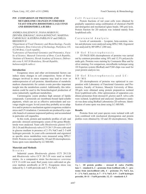
Fig. 1. 1D protein profiles – stressed r. rubra (NaOh);<br />
lane 1 – standard 6, 2 – proteome from lyofilized cells, 3 – proteome<br />
from non-lyofilized cells, 4 – proteome 2% NaCl, lyo,<br />
5 – 2 % NaCl, non-lyo, 6–7 – 5 % NaCl, 8–9 – 2 mM hydrogen<br />
peroxide, 10–11 – 5 mM peroxide; 12, 13 – standards 4, 5