3. FOOD ChEMISTRy & bIOTEChNOLOGy 3.1. Lectures
3. FOOD ChEMISTRy & bIOTEChNOLOGy 3.1. Lectures
3. FOOD ChEMISTRy & bIOTEChNOLOGy 3.1. Lectures

Create successful ePaper yourself
Turn your PDF publications into a flip-book with our unique Google optimized e-Paper software.
Chem. Listy, 102, s265–s1311 (2008) Food Chemistry & Biotechnology<br />
P39 TriCHOsPOrON CuTANeuM: CELL<br />
ADhESION ON CELLOPhANE SuRFACE<br />
JITKA HRDInOVá, TEREZA KRULIKOVSKá,<br />
VLADIMíR JIRKů, OLGA SCHREIBEROVá, ALEnA<br />
ČEJKOVá and JAn MASáK<br />
Institute of Chemical Technology Prague,<br />
Technická 5, 166 28 Praha 6 – Dejvice, Czech Republic,<br />
jitka.hrdinova@vscht.cz<br />
Introduction<br />
Recently we have faced the problem of increasing<br />
amount of solid cellulose wastes that come mainly from agriculture<br />
activities, food industry as well as from municipal<br />
waste 1,2 .<br />
Cellulose is considered to be a solid nontoxic pollutant;<br />
however, its recalcitrant nature causes many difficulties in<br />
removal of cellulosic waste. The microbial degradation is one<br />
of the possibilities how it could be solved.<br />
Colonization of solid material by microbial cells is<br />
the crucial step in their biodegradation. Biofilm formation<br />
by cellulolytic microorganisms is not investigated enough.<br />
Therefore, establishing more efficient arrangement for technological<br />
solubilization of solid cellulosic wastes could be<br />
achieved using cellulolytic biofilms, formed by direct colonization<br />
of these wastes by cell populations. In this association,<br />
cellophane was chosen as a representative of solid cellulosic<br />
substrates (carrier) and the yeast Trichosporon cutaneum as a<br />
relatively little researched cellulolytic strain.<br />
Experimental<br />
The yeast Trichosporon cutaneum CCY 30-4-5 was<br />
obtained from Department of Genetics and Microbiology,<br />
Faculty of Science, Charles University, Czech Republic.<br />
Inoculum was grown in complex medium and after<br />
2 days it was replaced in minimal medium supplemented<br />
with 1% cellulosic substrates as a sole source of carbon. Sigmacell<br />
Type 101, carboxymethylcellulose – CMC, hydroxyethylcellulose<br />
– HEC, cellophane and filter paper was used<br />
as the representatives of these substrates. Minimal medium<br />
composition in g dm –3 : KH 2 PO 4 – 1.7; na 2 HPO 4 . 7H2 O<br />
– 0.75; (nH 4 ) 2 SO 4 – 5.0; MgSO 4 . 7H2 O – 0.02; CaCl 2 – 0.02;<br />
FeSO 4 . 7H2 O – 0.001; MnSO 4 . H2 O – 0.001, pH 5.8. The<br />
temperature was maintained at 28 °C. Cultures were harvested<br />
during the exponential phase; at 48–72 hr. Separated<br />
and rinsed cells were used for experiments.<br />
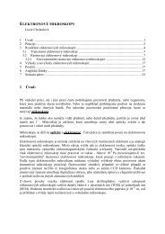
F C 8 1 F l o w C e l l D e s c r i p t i o n<br />
The FC 81 Flow Cell in Fig. 1. is a flat plate flow cell<br />
designed for use with transmitted light microscopes and it<br />
was used for observation of biofilm formation by yeast Trichosporon<br />
cutaneum. The capability of the yeast cells to<br />
colonize cellophane as well as the effect of shear stress was<br />
investigated.<br />
s654<br />
•<br />
•<br />
•<br />
•<br />
Experiment conditions:<br />
Cell suspension – OD 400 nm 0.1<br />
Temperature – 22 °C<br />
Flow rate – 2-20 ml min –1<br />
Time period – 2 hr.<br />
Fig. 1. Photograph of the FC 81 Flow Cell (biosurface Technologies,<br />
Corp., uSA)<br />
Experiments were carried out as shown by Fig. 2.;<br />
Fig. <strong>3.</strong> illustrates an emplacement of cellophane stripe in the<br />
flow cell.<br />
H y d r o p h o b i t y o f Y e a s t C e l l s<br />
The yeast populations were prepared in cultivation<br />
medium supplemented with different types of cellulosic substrates.<br />
Hydrophobity of yeast cell surface was determined<br />
using MATH method 3 .<br />
M i c r o s c o p y a n d I m a g e A n a l y s i s<br />
The colonization of cellophane was observed with<br />
transmitted light microscopy and analysis of the images was<br />
accomplished with Lucia (Laboratory Imaging, Ltd., UK).<br />
The areal parameters of objects (colonies, cells) such as<br />
length, width and colonized area were measured with image<br />
analysis4. The observation area of the flow cell was divided<br />
into three fields (inflow, middle, and outflow). Ten images<br />
were taken in every field.<br />
•<br />
•<br />
•<br />
Microscope – nIKOn Eclipse E400, Plan Fluor objective,<br />
10 ×/0.30, Ph1 DLL, ∞/0.17 WD 16.0 (Japan)<br />
Filter – 45 mm, nCB11<br />
Camera and software – Canon PowerShot A620, Zoom-<br />
Browser EX 5.5